Creatine
Aminoacids Metabolism

Author: Gianpiero Pescarmona
Date: 03/10/2011

Description

STRUCTURE

Pathway

FUNCTION

SUPPLY

SYNTHESIS

creatine biosynthesis

Creatine biosynthetic pathway. HA, h′epatic artery; PV, portal vein; HV, hepatic vein; SAM, S-adenosylmethionine; SAH, S-adenosylhomocysteine; GAMT, guanidinoacetate N-methyltransferase; AGAT, L-arginine:glycine amidinotransferase; GAA, guanidinoacetate acid.

Creatine synthesis: hepatic metabolism of guanidinoacetate and creatine in the rat in vitro and in vivo, 2009

DIET

CATABOLISM

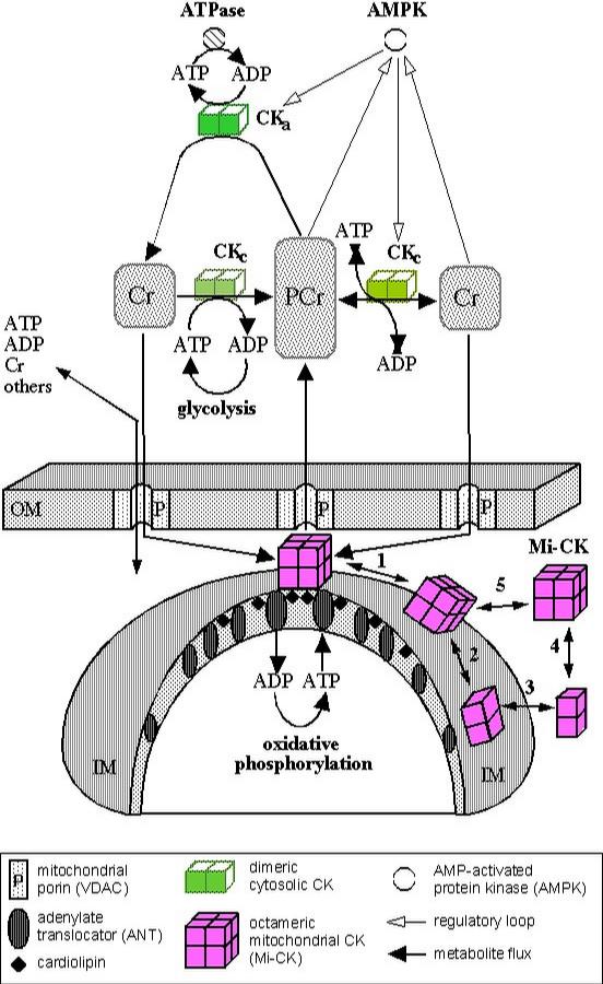
Multiple Functions of Creatine Kinase for Cellular Energetics: a Scientific Rationale for Creatine Supplementation

Impaired muscle uptake of creatine in spinal and bulbar muscular atrophy, 2016

Objective

The aim of this study was to explore the pathomechanism underlying the reduction of serum creatinine (Cr) concentrations in spinal and bulbar muscular atrophy (SBMA).
Methods

We evaluated blood chemistries, motor function, and muscle mass measured by dual‐energy X‐ray absorptiometry in male subjects with SBMA (n = 65), amyotrophic lateral sclerosis (ALS; n = 27), and healthy controls (n = 25). We also examined the intramuscular concentrations of creatine, a precursor of Cr, as well as the protein and mRNA expression levels of the creatine transporter (SLC6A8) in autopsy specimens derived from subjects who had SBMA and ALS and disease controls. Furthermore, we measured the mRNA expression levels of SLC6A8 in cultured muscle cells (C2C12) transfected with the polyglutamine‐expanded androgen receptor (AR‐97Q).
Results

Serum Cr concentrations were significantly lower in subjects with SBMA than in those with ALS (P < 0.001), despite similar muscle mass values. Intramuscular creatine concentrations were also lower in with the autopsied specimen of SBMA subjects than in those with ALS subjects (P = 0.018). Moreover, the protein and mRNA expression levels of muscle SLC6A8 were suppressed in subjects with SBMA. The mRNA levels of SLC6A8 were also suppressed in C2C12 cells bearing AR‐97Q.

Comments
2013-04-08T07:32:26 - Alessandra Caracciolo

CREATINE

Creatine is a nitrogenous organic acid that occurs naturally in vertebrates and helps to supply energy to all cells in the body, primarily muscle. This is achieved by increasing the formation of adenosine triphosphate (ATP). In solution, creatine is in equilibrium with creatinine.

Creatine is naturally produced in the human body from amino acids primarily in the kidney and liver. It is transported in the blood for use by muscles. Approximately 95% of the human body's total creatine is located in skeletal muscle. Creatine is not an essential nutrient, as it is manufactured in the human body from L-arginine, glycine, and L-methionine.
In humans and animals, approximately half of stored creatine originates from food (about 1 g/day, mainly from meat).

The enzyme GATM (L-arginine:glycine amidinotransferase (AGAT)) is a mitochondrial enzyme responsible for catalyzing the first rate-limiting step of creatine biosynthesis, and is primarily expressed in the kidneys and pancreas. The second enzyme in the pathway (GAMT, Guanidinoacetate N-methyltransferase) is primarily expressed in the liver and pancreas.

Creatine, synthesized in the liver and kidney, is transported through the blood and taken up by tissues with high energy demands, such as the brain and skeletal muscle, through an active transport system. The concentration of ATP in skeletal muscle is usually 2-5 mM, which would result in a muscle contraction of only a few seconds. Fortunately, during times of increased energy demands, the phosphagen (or ATP/PCr) system rapidly resynthesizes ATP from ADP with the use of phosphocreatine (PCr) through a reversible reaction with the enzyme creatine kinase (CK). Additionally, in most muscles, the ATP regeneration capacity of CK is very high and is therefore not a limiting factor. Although the cellular concentrations of ATP are small, changes are difficult to detect because ATP is continuously and efficiently replenished from the large pools of PCr and CK. Creatine has the ability to increase muscle stores of PCr, potentially increasing the muscle’s ability to resynthesize ATP from ADP to meet increased energy demands .

Creatine transport

Creatine transport into red blood cells : The permeation of creatine from plasma into normal human red blood cells was investigated by means of 1-[14C]-creatine. Two statistically different Vmax and Km values were found for lower and for higher creatine concentrations of the plasma, respectively, indicating two types of transport with different affinities and capacities. It is suggested that the high affinity process is an active transport, while the low affinity transport represents an exchange diffusion. There is little, if any, effect of pH in the range of 6.9-7.9 on the transport. The total creatine concentration of the red cells did not change significantly even with high creatine concentration of the plasma during 6 h incubation at 37 degrees C. The in vitro experiments showed a daily exchange of cellular creatine of 20%, the t0.5 being about 2.5 days. Creatine transport into red blood cells

Electrolysis stimulates creatine transport : Electrical field stimulation of isolated, incubated rodent skeletal muscles is a frequently used model to study the effects of contractions on muscle metabolism. In this study, this model was used to investigate the effects of electrically stimulated contractions on creatine transport. Soleus and extensor digitorum longus muscles of male NMRI mice were incubated in an oxygenated Krebs buffer between platinum electrodes. Muscles were exposed to [14C]creatine for 30 min after either 12 min of repeated tetanic isometric contractions (contractions) or electrical stimulation of only the buffer before incubation of the muscle (electrolysis). Electrolysis was also investigated in the presence of the reactive oxygen species (ROS) scavenging enzymes superoxide dismutase (SOD) and catalase. Both contractions and electrolysis stimulated creatine transport severalfold over basal. The amount of electrolysis, but not contractile activity, induced (determined) creatine transport stimulation. Incubation with SOD and catalase at 100 and 200 U/ml decreased electrolysis-induced creatine transport by ∼50 and ∼100%, respectively. The electrolysis effects on creatine uptake were completely inhibited by β-guanidino propionic acid, a competitive inhibitor of (creatine for) the creatine transporter (CRT), and were accompanied by increased cell surface expression of CRT. Muscle glucose transport was not affected by electrolysis. So, electrical field stimulation of incubated mouse muscles, independently of contractions per se, stimulates creatine transport by a mechanism that depends on electrolysis-induced formation of ROS in the incubation buffer. The increased creatine uptake is paralleled by an increased cell surface expression of the creatine transporter. Electrolysis stimulates creatine transport and transporter cell surface expression in incubated mouse skeletal muscle: potential role of ROS

Regulation of the creatine transporter by AMP-activated protein kinase in kidney epithelial cells :
The metabolic sensor AMP-activated protein kinase (AMPK) regulates several transport proteins, potentially coupling transport activity to cellular stress and energy levels. The creatine transporter (CRT; SLC6A8) mediates creatine uptake into several cell types, including kidney epithelial cells, where it has been proposed that CRT is important for reclamation of filtered creatine, a process critical for total body creatine homeostasis. Creatine and phosphocreatine provide an intracellular, high-energy phosphate-buffering system essential for maintaining ATP supply in tissues with high energy demands. Creatine uptake and apical surface biotinylation measurements in polarized S3 cells demonstrated parallel reductions in creatine influx and CRT apical membrane expression after AMPK activation with the AMP-mimetic compound 5-aminoimidazole-4-carboxamide-1-β-D-ribofuranoside. Moreover, AMPK may inhibit CRT indirectly via the mammalian target of rapamycin pathway.
AMPK inhibits apical membrane CRT expression in kidney proximal tubule cells, which could be important in reducing cellular energy expenditure and unnecessary creatine reabsorption under conditions of local and whole body metabolic stress. Regulation of the creatine transporter by AMP-activated protein kinase in kidney epithelial cells

USE AS A SUPPLEMENT AND AS A TREATMENT OF SOME DISEASES

Pharmacokinetics

Endogenous serum or plasma creatine concentrations in healthy adults are normally in a range of 2–12 mg/L. A single 5 g (5000 mg) oral dose in healthy adults results in a peak plasma creatine level of approximately 120 mg/L at 1–2 hours post-ingestion. Creatine has a fairly short elimination half-life, averaging just less than 3 hours, so to maintain an elevated plasma level it would be necessary to take small oral doses every 3–6 hours throughout the day. After the "loading dose" period (1–2 weeks, 12-24 g a day), it is no longer necessary to maintain a consistently high serum level of creatine. As with most supplements, each person has their own genetic "preset" amount of creatine they can hold. The rest is eliminated out of the body as waste. Creatine is consumed by the body fairly quickly, and if one wishes to maintain the high concentration of creatine, Post-loading dose, 2-5 g daily is the standard amount to intake.

Creatine has been demonstrated to cause modest increases in strength in people with a variety of neuromuscular disorders. Creatine supplementation has been, and continues to be, investigated as a possible therapeutic approach for the treatment of muscular, neuromuscular, neurological and neurodegenerative diseases (arthritis, congestive heart failure, Parkinson's disease, disuse atrophy, gyrate atrophy, McArdle's disease, Huntington's disease, miscellaneous neuromuscular diseases, mitochondrial diseases, muscular dystrophy, and neuroprotection), and depression.

Creatine supplements are athletic aids used to increase high-intensity athletic performance. There was once some concern that creatine supplementation could affect hydration status and heat tolerance and lead to muscle cramping and diarrhea, but recent studies have shown these concerns to be unfounded.
There are reports of kidney damage with creatine use, such as interstitial nephritis; patients with kidney disease should avoid use of this supplement. In similar manner, liver function may be altered, and caution is advised in those with underlying liver disease, although studies have shown little or no adverse impact on kidney or liver function from oral creatine supplementation. In 2004 the European Food Safety Authority (EFSA) published a record which stated that oral long-term intake of 3g pure creatine per day is risk-free .

Long-term administration of large quantities of creatine is reported to increase the production of formaldehyde, which has the potential to cause serious unwanted side-effects. However, this risk is largely theoretical because urinary excretion of formaldehyde, even under heavy creatine supplementation, does not exceed normal limits.

Extensive research has shown that oral creatine supplementation at a rate of 5 to 20 grams per day appears to be very safe and largely devoid of adverse side-effects, while at the same time effectively improving the physiological response to resistance exercise, increasing the maximal force production of muscles in both men and women.

A meta analysis performed in 2008 found that creatine treatment resulted in no abnormal renal, hepatic, cardiac or muscle function.

Wikipedia
Supplements
Creatine supplementations

AddThis Social Bookmark Button