the-benefits-of-n-acetylcysteine
diabete, dimagrimento,
GSH administration
Curcumin
Pulmonary protective effects of curcumin against paraquat toxicity. 2000
A single intraperitoneal injection of PQ (50 mg/kg) resulted in a significant rise in the levels of protein, angiotensin converting enzyme (ACE), alkaline phosphatase (AKP), N-acetyl-beta-D-glucosaminidase (NAG) and thiobarbituric acid reactive substances (TBARS), and neutrophils in the bronchoalveolar lavage fluid (BALF), while a decrease in glutathione levels.
Modulation of cyclophosphamide-induced early lung injury by curcumin, an anti-inflammatory antioxidant. 1995
Cyclophosphamide causes lung injury in rats through its ability to generate free radicals with subsequent endothelial and epithelial cell damage. In order to observe the protective effects of a potent anti-inflammatory antioxidant, curcumin (diferuloyl methane) on cyclophosphamide-induced early lung injury, healthy, pathogen free male Wistar rats were exposed to 20 mg/100 g body weight of cyclophosphamide, intraperitoneally as a single injection. Prior to cyclophosphamide intoxication oral administration of curcumin was performed daily for 7 days. At various time intervals (2, 3, 5 and 7 days post insult) serum and lung samples were analyzed for angiotensin converting enzyme, lipid peroxidation, reduced glutathione and ascorbic acid. Bronchoalveolar lavage fluid was analyzed for biochemical constituents. The lavage cells were examined for lipid peroxidation and glutathione content. Excised lungs were analyzed for antioxidant enzyme levels. Biochemical analyses revealed time course increases in lavage fluid total protein, albumin, angiotensin converting enzyme (ACE), lactate dehydrogenase, N-acetyl-beta-D-glucosaminidase, alkaline phosphatase, acid phosphatase, lipid peroxide levels and decreased levels of glutathione (GSH) and ascorbic acid 2, 3, 5 and 7 days after cyclophosphamide intoxication. Increased levels of lipid peroxidation and decreased levels of glutathione and ascorbic acid were seen in serum, lung tissue and lavage cells of cyclophosphamide groups. Serum angiotensin converting enzyme activity increased which coincided with the decrease in lung tissue levels. Activities of antioxidant enzymes were reduced with time in the lungs of cyclophosphamide groups.(ABSTRACT TRUNCATED AT 250 WORDS)
Losartan
Enalapril and captopril enhance glutathione-dependent antioxidant defenses in mouse tissues. 2000
These findings support our previous reports on the enalapril- and captopril-induced enhancement of endogenous antioxidant defenses and include new data on glutathione-dependent defenses, thus furthering current knowledge on the association of ACE inhibition and antioxidants.
The effects of losartan and enalapril therapies on the levels of nitric oxide, malondialdehyde, and glutathione in patients with essential hypertension. 2002
Several recent studies have shown that essential hypertension is associated with increased oxidative stress, which may cause hypertension via enhanced oxidation and inactivation of nitric oxide. In this study, we investigated the malondialdehyde, nitric oxide, and glutathione levels in newly diagnosed essential hypertensive patients and whether or not there was any effect of antihypertensive treatment with angiotensin II type 1 receptor antagonist, losartan or angiotensin converting enzyme inhibitor, enalapril on plasma malondialdehyde, nitric oxide, and glutathione values. We selected 17 patients (F/M: 10/7, mean age: 46.12 +/- 9.2 years) for enalapril therapy (10-20 mg/d) and 14 patients (F/M: 8/6, mean age: 47.7 +/- 7.5 years) for losartan therapy (50-100 mg/d), and compared them with 12 normotensive controls. At the beginning of the study, both treated groups showed significantly higher plasma malondialdehyde and lower glutathione and nitric oxide in exhaled air compared to the control group. After 9 weeks of enalapril and losartan treatment, both systolic and diastolic pressure were significantly reduced. Both enalapril and losartan produced a significant decrease in plasma malondialdehyde and a significant increase in plasma glutathione levels and nitric oxide in exhaled air after 9 weeks. Initial values of plasma nitrate levels in patient groups were similar to the control group and increased significantly after the treatment period. In conclusion, both losartan and enalapril may be regulators between oxidant stress and the antioxidant system.
Glycine and NAC (N-acetylcysteine) administration
 aging and glutathione
Deficient Synthesis of Glutathione Underlies Oxidative Stress in Aging and Can Be Corrected by Dietary Cysteine and Glycine Supplementation, 2011
- Abstract
Background: Aging is associated with oxidative stress, but underlying mechanisms remain poorly understood.
Objective: We tested whether glutathione deficiency occurs because of diminished synthesis and contributes to oxidative stress in aging and whether stimulating glutathione synthesis with its precursors cysteine and glycine could alleviate oxidative stress.
Design: Eight elderly and 8 younger subjects received stable-isotope infusions of [2H(2)]glycine, after which red blood cell (RBC) glutathione synthesis and concentrations, plasma oxidative stress, and markers of oxidant damage (eg, F(2)-isoprostanes) were measured. Elderly subjects were restudied after 2 wk of glutathione precursor supplementation.
Results: Compared with younger control subjects, elderly subjects had markedly lower RBC concentrations of glycine (486.7 ± 28.3 compared with 218.0 ± 23.7 μmol/L; P < 0.01), cysteine (26.2 ± 1.4 compared with 19.8 ± 1.3 μmol/L; P < 0.05), and glutathione (2.08 ± 0.12 compared with 1.12 ± 0.18 mmol/L RBCs; P < 0.05); lower glutathione fractional (83.14 ± 6.43% compared with 45.80 ± 5.69%/d; P < 0.01) and absolute (1.73 ± 0.16 compared with 0.55 ± 0.12 mmol/L RBCs per day; P < 0.01) synthesis rates; and higher plasma oxidative stress (304 ± 16 compared with 346 ± 20 Carratelli units; P < 0.05) and plasma F(2)-isoprostanes (97.7 ± 8.3 compared with 136.3 ± 11.3 pg/mL; P < 0.05). Precursor supplementation in elderly subjects led to a 94.6% higher glutathione concentration, a 78.8% higher fractional synthesis rate, a 230.9% higher absolute synthesis rate, and significantly lower plasma oxidative stress and F(2)-isoprostanes. No differences in these measures were observed between younger subjects and supplemented elderly subjects.

Mean (±SEM) glutathione (GSH) concentrations (A), GSH fractional synthesis rates (B), and GSH absolute synthesis rates © in young control (n = 8) and elderly (n = 8) subjects before and after supplementation with cysteine or glycine. Values are significantly different at P < 0.05 on the basis of 1-factor ANOVA and Bonferroni-corrected t-tests. A: a Significantly different from the elderly before supplementation, P < 0.05; b not significantly different from the elderly after supplementation; c significantly different from the elderly before supplementation, P < 0.05. B: a significantly different from the elderly before supplementation, P < 0.01; b not significantly different from the elderly after supplementation; c significantly different from the elderly after supplementation, P < 0.01. C: a significantly different from the elderly before supplementation, P < 0.01; b not significantly different from the elderly after supplementation; c significantly different from the elderly after supplementation, P < 0.01. RBC, red blood cells.
Conclusions: Glutathione deficiency in elderly humans occurs because of a marked reduction in synthesis. Dietary supplementation with the glutathione precursors cysteine and glycine fully restores glutathione synthesis and concentrations and lowers levels of oxidative stress and oxidant damages. These findings suggest a practical and effective approach to decreasing oxidative stress in aging.
Tesi 2015
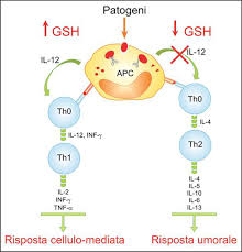
Utilizzo di molecole pro-glutatione per modulare la risposta immunitaria Th1/Th2 durante le infezioni virali, 2015
glutathione+fraternale
brundu+fraternale+glutathione
Dexamethasone improves redox state in ataxia telangiectasia cells by promoting an NRF2‐mediated antioxidant response, 2016
- Ataxia telangiectasia (A‐T) is a rare incurable neurodegenerative disease caused by biallelic mutations in the gene for ataxia‐telangiectasia mutated (ATM). The lack of a functional ATM kinase leads to a pleiotropic phenotype, and oxidative stress is considered to have a crucial role in the complex physiopathology. Recently, steroids have been shown to reduce the neurological symptoms of the disease, although the molecular mechanism of this effect is largely unknown. In the present study, we have demonstrated that dexamethasone treatment of A‐T lymphoblastoid cells increases the content of two of the most abundant antioxidants [glutathione (GSH) and NADPH] by up to 30%. Dexamethasone promoted the nuclear accumulation of the transcription factor nuclear factor (erythroid‐derived 2)‐like 2 to drive expression of antioxidant pathways involved in GSH synthesis and NADPH production. The latter effect was via glucose 6‐phosphate dehydrogenase activation, as confirmed by increased enzyme activity and enhancement of the pentose phosphate pathway rate. This evidence indicates that glucocorticoids are able to potentiate antioxidant defenses to counteract oxidative stress in ataxia telangiectasia, and also reveals an unexpected role for dexamethasone in redox homeostasis and cellular antioxidant activity.
Glutathione increase by the n‐butanoyl glutathione derivative (GSH‐C4) inhibits viral replication and induces a predominant Th1 immune profile in old mice infected with influenza virus
Oxidation-induced increase in activity of angiotensin converting enzyme in the rat kidney. 1988
The activity of ACE was also increased by the diamide-pretreatment of the isolated membrane fraction of the renal cortex, thereby indicating that the increase in activity was not due to oxidation of endogenous glutathione (GSH) that may lower the ACE activity, but rather that ACE itself was oxidized.
Ginkgo Biloba Extract (EGb 761) Normalizes Hypertension in 2K, 1C Hypertensive Rats: Role of Antioxidant Mechanisms, ACE Inhibiting Activity and Improvement of Endothelial Dysfunction, 2011
Abstract
The 2 kidney, 1-clip (2K, 1C) model of hypertension was used to investigate the potential antihypertensive effect of a standardized leaf extract of Ginkgo biloba (EGb 761). Clipping of the renal artery resulted in gradual elevation of the systolic blood pressure (SBP) reaching a plateau after 4 weeks of surgery. Treatment of hypertensive rats with EGb 761 (60, 90, 180 mg/kg/day orally) was therefore started 4 weeks after surgery and continued for 3 weeks. This led to a dose-dependent reduction in SBP with no significant change in heart rate. Control hypertensive rats showed a significant elevation of total protein thiols (Pr-SHs level) in both clipped and non-clipped kidneys as well as in the serum. However, glutathione peroxidase (GSH-Px) activity was decreased in the clipped kidneys but elevated in the non-clipped ones and in the blood. The malondialdehyde (MDA) level was raised in clipped kidneys but not in non-clipped ones nor in the serum. Nitric oxide (NO level) and angiotensin converting enzyme (ACE) activity were increased in both clipped and non-clipped kidneys but not in the serum. Endothelium-dependent and -independent relaxation of aortic rings towards acetylcholine (Ach) and sodium nitroprusside (SNP) were impaired. Treatment with EGb 761 (180 mg/kg/day for 3 weeks) was associated with recovery of GSH-Px activity in clipped kidneys, inhibition of ACE activity in both kidneys and a reduction in the elevated NO level of the non-clipped kidneys, decreased responsiveness to the vasoconstrictor NE and improvement of endothelial function as evidenced by restoration of endothelium-dependent vasorelaxation induced by Ach. The observed beneficial effects of the EGb 761 may be attributed to different factors, including ACE inhibition and maintenance of cellular antioxidant capacity as well as preserving vascular reactivity towards endothelium-dependent and -independent vasodilators while inhibiting responses to vasoconstrictor.
A Review of Dietary (Phyto)Nutrients for Glutathione Support, 2019