Immunoglobulin G Class (IgG)
Immunoglobulins

Author: matteo rosso
Date: 02/04/2009

Description

Immunoglobulins G, of which there are four subclasses (IgG1, IgG2, IgG3 and IgG4) are the quantitatively class of antibody more represented in the serum, but are also present in the extravascular environement.
They are produced during the secondary response and activate the classical way of complement (interaction of C1, C4 and C2 with the antigen-antibody complex). IgGs play a role during the opsonisation of microorganisms encouraging their phagocytosis by specific receptors (FcgRI) for the Fc fragment of IgG expressed on the cell membranes of neutrophils and macrophages.

IgG function

The binding of IgG on the surface of foreign cells increases the efficiency of cell lysis (ADCC mechanism: antibody-dependent cell-mediated cytotoxicity) by NK cells, using a low-affinity receptor expressed by these cells: the FcgRIII.
Other low-affinity receptors (FcgRII) are expressed by monocytes, macrophages, neutrophils, eosinophils, platelets and B lymphocytes. These receptors facilitate the removal of circulating immune complexes from cells engulf.

Fc-gR : receptor for Fc fragment of IgG

FcgR belong to the superfamily of immunoglobulins and plays a vital role in the phagocytosis of opsonised microorganisms.
The family of Fc-gR includes many subtypes, such as FcγRI (CD64), FcγRIIA (CD32), FcγRIIB (CD32), FcγRIIIA (CD16a) and FcgRIIIB (CD16b), each of which differs from the others by a different affinity towards antibodies, inequality due to a different molecular structure.
FcγRI binds more strongly than IgG they do FcgRII and FcgRIII. This increased affinity is due to an extracellular portion composed of three Ig-like domains, one in more than FcgRII and FcgRIII. This property allows the activation of FcgRI by one molecule of IgG without which an immune complex can not be activated.
This class of receptors has other features: the structure is similar to that of MHC class I and is involved in antibody transfer from mother to fetus during pregnancy and after during lactation (FcRn: neonatal Fc receptor).

Fc-gR modulation by Genistein

Kawasaki disease and soy: potential role for isoflavone interaction with Fcγ receptors, 2013

Kawasaki disease is a diffuse vasculitis occurring in children and showing predilection for the coronary arteries. Differences in diet between Asians and Westerners are touted as reasons for certain ethnic-related discrepancies in susceptibility to cardiovascular disease and cancer in adults. Surprisingly, these cultural dietary differences have not been previously considered as the source of the discrepancy in KD incidence among these ethnic populations. Recent data from genetic studies have highlighted the role of specific immune receptors in the pathogenesis of KD. Functions of the Fcγ receptors (FcGRs) are modulated by isoflavones in soy, in particular, genistein. Epidemiological data from Hawaiian populations support an association between soy consumption and KD. These observations form the basis of a hypothesis: isoflavones participate in KD pathogenesis by modulating function of the FcGRs and by disrupting the balance between activation and inhibition of the inflammatory response.

Fc receptors: Cell activators of antibody functions, 2013

IgG molecules bind to their cognate antigens and are in turn recognized by specific receptors (Fcγ receptors) on the membrane of leukocytes. Crosslinking these receptors on the surface of leukocytes leads to activation of several effector cell functions. These effector functions are geared toward the destruction of microbial pathogens and the induction of an inflammatory state that is beneficial during infections. However, in autoimmune diseases, antibodies can direct these effector functions against normal tissues and cause severe tissue damage. In recent years, several factors that can modulate the IgG-FcγR interaction have been elucidated. In this review, we describe the main types of Fcγ receptors, and our current view of how antibody variants interact with these receptors to initiate different cell responses.

Mechanism of degradation of monoclonal antibodies. FcgR, Fc gamma receptor; FcRn, Brambell receptor.

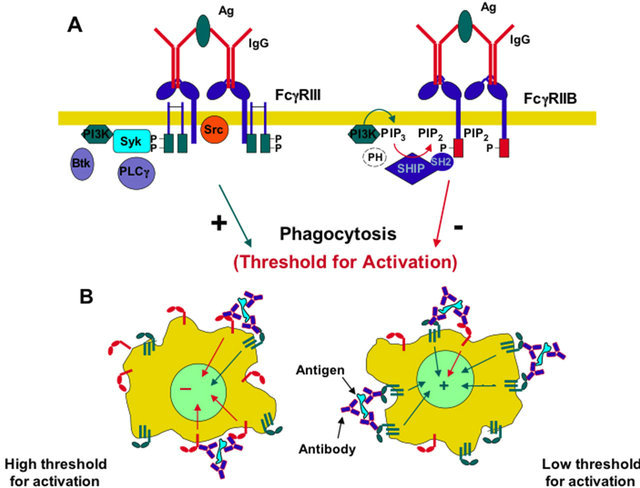
FcgR generate signals through an activator site called ITAM (Immunoreceptor tyrosine-based activation motif). Each ITAM is composed by a pattern of amino acids (YXXL) in succession in the queue dual intracellular receptor. When thyroxines (Y) are added to the phosphate groups by action of specific tyrosine kinases, the signal is transmitted within the cell. This phosphorylation always coupling the Fc receptor with its specific ligand.
ITAM stimulates phagocytosis in macrophages. FcgRI and FcgRIIIA lack of ITAM but still transmit the signal using another protein function. This molecule, called FCG subunit contains two patterns (YXXL) characteristic of the ITAM, which, in effect, override the role played by this molecule.

These two patterns must be present in two copies, because one of the two (ITIM) seems to have inhibitory effects against phagocytosis. The inhibition is regulated by two enzymes, SHP-1 and SHP-2 that removes phosphate groups from tyrosine residues, thus blocking the positive response to the stimulus (Figure 2).

FCR recognizes microorganisms that have been linked by Abs (Figure 3). The interaction between antibodies bound to the surface of the cell and FCR actives cell of the immune system to destroy the target.

Figure 3: The picture depicts the action of macrophages against a microorganism coated with antibodies"

IgG synthesis

Signaling Proteins and Transcription Factors in Normal and Malignant Early B Cell Development, 2011

Figure 1: B cell development in adult mammals starts in bone marrow with the commitment of hematopoietic stem cells (HSCs) to the B cell lineage and ends with formation of mature B cells in peripheral secondary lymphoid organs (e.g., the spleen). It is the sequential expression and assembly of the components of the B cell antigen receptor (BCR) what defines each developmental stage. The first stage exhibiting commitment to the B cell lineage is the proB, and here the immunoglobulin heavy chain is in the process of recombination, and the signaling proteins Igα and Igβ are in surface forming complexes with chaperon proteins like calnexin (the proBCR). The next developmental stage, the preB, happens after the heavy chain was successfully recombined and the preBCR is assembled. In this stage, the light chain is recombined and unrearranged heavy chain alleles are excluded. After light chain recombination and pairing with the heavy chain and Igα and Igβ the mature BCR is formed, the B cell is in the immature (in bone marrow) and transitional (in periphery) stages. Here, B cell mechanisms of self-tolerance are active allowing self- and nonself-recognition by the mature B cell. Transition to the mature stage happens if the BCR of the immature/transitional B cell does not find its cognate antigen after several days of bone marrow and peripheral trafficking.

Figure 2: (a) The Ig heavy and light chain genes are comprised of constant and variable regions, where the variable region is formed by an n number of segments termed V (variable), D (diversity), and J (joining) in the heavy chain and by segments V and J in the light chain. These segments are brought together by a site-specific recombination process termed VDJ recombination responsible for the extensive repertoire of BCR specificities. There are two loci for light chain, κ and λ. Here, all the loci are shown in germline configuration, previous to the process of VDJ recombination. (b) The early stages of B cell development are differentiated by the process of VDJ recombination, and the heavy (IgH) and light (IgL) chains are recombined in the proB and preB stages, respectively. Each stage is further subdivided according to the sequential assembly of the VDJ segments. Replication and recombination processes are mutually exclusive as denoted by the circular arrows and VDJ signs inside the cell. Dashed lines separating proB and preB stages indicate checkpoints where signaling from the preBCR and BCR is required for positive selection and progression along the B cell maturation pathway. Continuous lines indicate the main receptors controlling each developmental stage. The differential intensity in the IL-7 green line indicates the sub-stages where a higher or lower concentration of the IL-7R ligand is required.

Figure 3: Homeostatic and leukemic expression of receptors, signaling proteins, and transcription factors along the B cell pathway. Developmental stages are indicated starting with the hematopoietic stem cell (HSC), the common lymphoid progenitor (CLP), and into the B cell pathway, stages proB, preB, and mature B cells. Normal gene expression along the developmental pathway is indicated with blue bars, and expression dependency between proteins is indicated with arrows. Most common modified forms of these receptors, signaling proteins, and transcription factors associated with acute lymphoblastic leukemia are also indicated.

Free immunoglobulin light chain: Its biology and implications in diseases 2011 Fulltext

additional links

Molecular features responsible for the absence of immunoglobulin heavy chain protein synthesis in an IgH− subgroup of multiple myeloma 2000

Synthesis and secretion of light-chain immunoglobulin in two successive cycles of synchronized plasmacytoma cells, 1976

Biology of immunoglobulin light chains

Immunohistochemical staining of κ producing bone marrow plasma cells from a patient with MM using fluorescein-conjugated, anti-κ antiserum. 5 plasma cells can be seen. B. Diagrammatic representation of plasma cells producing intact immunoglobulins with monomeric κ and dimeric λ FLC molecules.

Plasma cells produce one of five heavy chain types, together with κ or λ molecules. There is approximately 40% excess FLC production over heavy chain synthesis, to allow proper conformation of the intact immunoglobulin molecules. As mentioned previously, there are twice as many κ-producing plasma cells as λ-producing plasma cells. κ FLCs are normally monomeric, while λ FLCs tend to be dimeric, joined by disulphide bonds, however higher polymeric forms of both FLCs may occur

Nephron showing filtration, metabolism and excretion of FLCs.

Because of the huge metabolic capacity of the proximal tubule, the amount of FLCs in urine, even when production is considerably increased, is more dependent upon renal function than synthesis by the tumour. As a consequence, serum and urine FLC concentrations may not be similar during the evolution of LCMM. This is shown in a hypothetical patient in Figure 3.8. The red line shows the steady increase in sFLCs as the tumour grows over the first 12 months. When synthesis of FLCs exceeds 10-30g/day (greater than 30 times normal) there is an overflow proteinuria and large amounts of FLCs enter the urine. It is normally at this point that patients with LCMM are identified.

Co-translational modification of nascent immunoglobulin heavy and light chains., 1979

The nucleotide sequence of a methionine tRNA which functions in protein elongation in mouse myeloma cells. 1975

IgG synthesis regulation

Liver-X-receptor activator prevents homocysteine-induced production of IgG antibodies from murine B lymphocytes via the ROS-NF-kappaB pathway. 2007

  • Our previous study showed that homocysteine (Hcy) promotes proliferation of mouse splenic B lymphocytes. In this study, we investigated whether Hcy could stimulate the production of IgG antibodies. Hcy significantly increased the production of IgG antibodies from resting B lymphocytes. B lymphocytes from ApoE-knockout mice with hyperhomocysteinemia showed elevated IgG secretion at either the basal Hcy level or in response to lipopolysaccharide. Hcy promoted reactive oxygen species (ROS) formation, and free radical scavengers, MnTMPyP decreased Hcy-induced IgG secretion. The inhibitor of NF-kappaB (MG132) also significantly reduced Hcy-induced IgG secretion. Furthermore, Hcy-induced formation of ROS, activation of NF-kappaB, and secretion of IgG could be inhibited by the liver-X-receptor (LXR) agonist T0901317. Thus, our data provide strong evidence that HHcy induces IgG production from murine splenic B lymphocytes both in vitro and in vivo. The mechanism might be through the ROS-NF-kappaB pathway and can be attenuated by the activation of LXR.

Down Syndrome: low Methionine

down syndrome Igg

Neonates with Downs syndrome but not other chromosomal abnormalities are associated with a deficiency of IgG.

DNA Methylation

Localized DNA Demethylation at Recombination Intermediates during Immunoglobulin Heavy Chain Gene Assembly, 2013

CHAPTER 83 FUNCTIONS OF B LYMPHOCYTES AND PLASMA CELLS IN IMMUNOGLOBULIN PRODUCTION, Williams Hematology

Free immunoglobulin light chain: Its biology and implications in diseases, 2011

  • Immunoglobulin light chain (IgLC) is a component of antibodies, but its free form is observed in the circulation, which originates from 10 to 40% excess synthesis over heavy chain in B cells. Complete antibodies function as a defined tetramer structure unit, H2L2; thus, separation of heavy and light chains results in considerable or complete loss of antigen-binding ability. Free IgLC has been considered as an inconsequential spillover during antibody assembly because, unlike heavy chain, neither effector functions such as complement activation nor specific-receptor binding has been identified in IgLCs. Free IgLC in sera and cerebrospinal fluids increases in inflammatory diseases such as autoimmune diseases and infections, presumably as a result of B-cell activation. This may be just a concomitant event during elevated disease activity, but recent findings suggest that free IgLC is involved in a wide range of immunological phenomena as a signaling effector or an anti-inflammatory molecule. These effects are likely to be intrinsic to IgLC. In this review, we attempt to give a comprehensive view about the biological roles of free IgLC together with the gene expression, secretion, antigen-binding ability, and its metabolic characteristics.

Fulltext

Comments
2014-05-01T11:47:08 - Gianpiero Pescarmona

BiP and Immunoglobulin Light Chain Cooperate to Control the Folding of Heavy Chain and Ensure the Fidelity of Immunoglobulin Assembly, 1999

  • The immunoglobulin (Ig) molecule is composed of two identical heavy chains and two identical light chains (H2L2). Transport of this heteromeric complex is dependent on the correct assembly of the component parts, which is controlled, in part, by the association of incompletely assembled Ig heavy chains with the endoplasmic reticulum (ER) chaperone, BiP. Although other heavy chain-constant domains interact transiently with BiP, in the absence of light chain synthesis, BiP binds stably to the first constant domain (CH1) of the heavy chain, causing it to be retained in the ER. Using a simplified two-domain Ig heavy chain (VH-CH1), we have determined why BiP remains bound to free heavy chains and how light chains facilitate their transport. We found that in the absence of light chain expression, the CH1 domain neither folds nor forms its intradomain disulfide bond and therefore remains a substrate for BiP. In vivo, light chains are required to facilitate both the folding of the CH1 domain and the release of BiP. In contrast, the addition of ATP to isolated BiP–heavy chain complexes in vitro causes the release of BiP and allows the CH1 domain to fold in the absence of light chains. Therefore, light chains are not intrinsically essential for CH1 domain folding, but play a critical role in removing BiP from the CH1 domain, thereby allowing it to fold and Ig assembly to proceed. These data suggest that the assembly of multimeric protein complexes in the ER is not strictly dependent on the proper folding of individual subunits; rather, assembly can drive the complete folding of protein subunits.

Co-translational modification of nascent immunoglobulin heavy and light chains, 2004

  • We have investigated the in vivo co-translational covalent modification of nascent immunoglobulin heavy and light chains. Nascent polypeptides were separated from completed polypeptides by ion-exchange chromatography of solubilized ribosomes on QAE-Sephadex. First, we have demonstrated that MPC 11 nascent heavy chains are quantitatively glycosylated very soon after the asparaginyl acceptor site passes through the membrane into the cisterna of the rough endoplasmic reticulum. Nonglycosylated completed heavy chains of various classes cannot be glycosylated after release from the ribosome, due either to rapid intramolecular folding and/or intermolecular assembly, which cause the acceptor site to become unavailable for the glycosylation enzyme. Second, we have shown that the formation of the correct intrachain disulfide loop within the first light chain domain occurs rapidly and quantitatively as soon as the appropriate cysteine residues of the nascent light chain pass through the membrane into the cisterna of the endoplasmic reticulum. The intrachain disulfide loop in the second or constant region domain of the light chain is not formed on nascent chains, because one of the cysteine residues involved in this disulfide bond does not pass through the endoplasmic reticulum membrane prior to chain completion and release from the ribosome. Third, we have demonstrated that some of the initial covalent assembly (formation of interchain disulfide bonds) occurs on nascent heavy chains prior to their release from the ribosome. The results are consistent with the pathway of covalent assembly of the cell line, in that completed light chains are assembled onto nascent heavy chains in MPC 11 cells (IgG2b), where a heavy-light half molecule is the major initial covalent intermediate; and completed heavy chains are assembled onto nascent heavy chains in MOPC 21 cells (IgG1), where a heavy chain dimer is the major initial disulfide linked intermediate.

Association of transport-defective light chains with immunoglobulin heavy chain binding protein, 1990

  • Immunoglobulin light chains are usually secreted from cells when they are synthesized alone or in molar excess of heavy chains, but, there have been reports of nonsecreted light chains. We wished to determine whether immunoglobulin heavy chain binding protein (BiP), which blocks the transport of free heavy chains, might be responsible for the lack of secretion of some light chains. In two murine lymphoid cell lines that synthesize but do not secrete immunoglobulin light chains, the free light chain polymers were found bound to BiP. Examination of 20 other cell lines and hybridomas failed to disclose any cells synthesizing free or excess light chains that associated with BiP; in all cases the free light chains were secreted as dimers. Despite their association with BiP and their blocked secretion, the aberrant light chains could combine with heavy chains and could be secreted as intact Ig molecules. Thus, while light chains do not usually express signals which allow them to bind to BiP, it appears that such signals can be expressed on certain light chains, resulting in their combination with BiP and blocked secretion. When single chain mutant cell lines are isolated from parental lines producing both heavy and light chains, they are almost always light chain producers suggesting that free heavy chains are much more toxic than free light chains. In both PC700 and P3X63Ag cells, however, clones that have lost either heavy chains or transport-defective light chains are present at the same frequency. Our findings that the light chains in both of these lines are associated with BiP raise the possibility that BiP actually contributes to heavy chain toxicity instead of preventing it.
2014-04-20T09:24:55 - Gianpiero Pescarmona

Assembly and Secretion of Heavy Chains that Do Not Associate Posttranslationally with Immunoglobulin Heavy Chain-binding Protein, 1987

  • Abstract. Heavy chain-binding protein (BiP) associates posttranslationally with nascent Ig heavy chains in the endoplasmic reticulum (ER) and remains associated with these heavy chains until they assemble with light chains.

The heavy chain-BiP complex can i
be precipitated by antibody reagents against either
component. To identify sites on heavy chain molecules
that are important for association with BiP, we have
examined 30 mouse myelomas and hybridomas that
synthesize Ig heavy chains with well characterized de-
letions. Mutant Ig heavy chains that *lack the C,1 do-
main could not be demonstrated to associate with BiP,*
whereas mutant Ig heavy chains with deletions of the
CH2 or CH3 domain were still able to associate with
BiP. In two light chain negative cell lines that pro-
duced heavy chains with deletions of the CH1 domain,
free heavy chains were secreted. When Ig assembly
and secretion were examined in mutants that did not
associate with BiP, and were compared with normal
parental lines, it was found that the rate of Ig secretion
was increased in the mutant lines and that the Ig
molecules were secreted in various stages of assembly.
In one mutant line (CH1-) approximately one-third of
the secreted Ig molecules were incompletely assem-
bled, whereas the Ig molecules secreted by the paren-
tal line were completely assembled. Our data show the
CH1 domain to be important for association with BiP
and that when this association does not occur, incom-
pletely assembled heavy chains can be secreted. This
implies a role for BiP in preventing the transport of
unassembled Ig molecules from the ER.
I MMUNOGLOBULIN biosynthesis, assembly, and trans-
port have been well characterized using both normal and
malignant lymphoid cells (7, 27).

The Ig molecule is composed of two identical heavy chain proteins and two identical light chain (LC) t proteins which are joined by interchain disulfide bonds. Nascent heavy and light chains contain a hydrophobic amino-terminal signal sequence which allows them to be cotranslationally translocated into the lumen of the endoplasmic reticulum (ER)

The addition of mannose core sugars to heavy chains occurs during this translocation
Very soon after translocation, heavy and light chains begin to assemble in the ER and interchain disulfide bonds are formed.

The sequence of subunit assembly appears to be determined by the heavy chain isotype.

  • IgM and some IgG2b assemble as heavy and light chain (HL) molecules and then H2~molecules,
  • whereas IgGt and IgGz, first form H2 molecules, then H2L, and finally intact H2L2
    molecules

Once assembly is complete, the Ig molecule is transported to the Golgi complex for further processing

The mechanism for directing the Ig molecule to Golgi is not well understood. Data
gathered from studying the transport of other proteins to the
Golgi complex suggest the existence of inherent transport
signals on protein molecules which direct their transport to
the Golgi complex (10, 20). It is not clear if transport se-
quences are present on the heavy chain, the light chain, or
both. After processing in the Golgi complex, the Ig molecule
is packaged into vesicles that are targeted to the plasma mem-
brane for secretion.
Usually, only completely assembled Ig molecules are
secreted. When assembly can not occur due to the absence
of light chain synthesis or is inhibited due to the under-
glycosylation of heavy chains in the case of IgM, the free
heavy chains are not transported to the Golgi complex and
are degraded internally (13, 17). A notable exception to this
occurs in lymphoproliferative heavy chain disease (HCD).
This disorder is characterized by the production and secre-
tion of heavy chains in the absence of LC production (11).
The secreted heavy chains are abnormal in that they contain
large protein deletions that usually involve the first constant
region domain (CH1) and occasionally the variable region

GRP78_HUMAN = BiP

iP and Immunoglobulin Light Chain Cooperate to Control the Folding of Heavy Chain and Ensure the Fidelity of Immunoglobulin Assembly, 1999

  • The immunoglobulin (Ig) molecule is composed of two identical heavy chains and two identical light chains (H2L2). Transport of this heteromeric complex is dependent on the correct assembly of the component parts, which is controlled, in part, by the association of incompletely assembled Ig heavy chains with the endoplasmic reticulum (ER) chaperone, BiP. Although other heavy chain-constant domains interact transiently with BiP, in the absence of light chain synthesis, BiP binds stably to the first constant domain (CH1) of the heavy chain, causing it to be retained in the ER. Using a simplified two-domain Ig heavy chain (VH-CH1), we have determined why BiP remains bound to free heavy chains and how light chains facilitate their transport. We found that in the absence of light chain expression, the CH1 domain neither folds nor forms its intradomain disulfide bond and therefore remains a substrate for BiP. In vivo, light chains are required to facilitate both the folding of the CH1 domain and the release of BiP. In contrast, the addition of ATP to isolated BiP–heavy chain complexes in vitro causes the release of BiP and allows the CH1 domain to fold in the absence of light chains. Therefore, light chains are not intrinsically essential for CH1 domain folding, but play a critical role in removing BiP from the CH1 domain, thereby allowing it to fold and Ig assembly to proceed. These data suggest that the assembly of multimeric protein complexes in the ER is not strictly dependent on the proper folding of individual subunits; rather, assembly can drive the complete folding of protein subunits.

Biology of immunoglobulin light chains

b+lymphocytes+grp78

Phosphorylation of eIF2α attenuates statin-induced apoptosis by inhibiting the stabilization and translocation of p53 to the mitochondria. 2013

In multiple myeloma cells, lovastatin has been shown to induce BiP/Grp78 and CHOP protein expression, the phosphorylation of eIF2α and apoptosis, as well as the cleavage of poly(ADP-ribose) polymerase (PARP).

The differential expressions of 78-kDa glucose-regulated protein of infiltrating plasma cells in peripheral joints with the histopathological variants of rheumatoid synovitis, 2009

The local production of pathogenic autoantibodies by plasma cells in synovium is one of the hallmarks of rheumatoid arthritis (RA). There may be a potential link between ectopic lymphoid neogenesis and the local autoimmunity in rheumatoid synovium. The unfolded protein response (UPR) has key roles in the development and maintenance of plasma cells secreting immunoglobulin. This study was designed to explore the potential links between the activation of the UPR of infiltrating plasma cells in inflamed peripheral joints and the histopathological variants of rheumatoid synovitis as well as the local production of pathogenic autoantibodies.

Methods

The variants of rheumatoid synovium were histopathologically classified into follicular and diffuse synovitis. Immunohistochemical and double-immunofluorescent stainings were performed to detect the expression of 78-kDa glucose-regulated protein (GRP78), a marker of activation of the UPR, in infiltrating plasma cells of synovium, and flow cytometry and immunoblotting analyses were performed to quantify GRP78 in plasma cells of synovial fluid in inflamed peripheral joints of RA. The detections were also taken in osteoarthritis (OA) as controls. The synovial fluid levels of anti-cyclic citrullinated peptide antibodies (anti-CCP) (IgG) were quantified with the enzyme-linked immunosorbent assay and corrected to those of total IgG in RA.

Results

Expressions of GRP78 were more intensive in infiltrating plasma cells in RA synovium relative to those in OA synovium (P < 0.001) and in synovium with follicular synovitis relative to that with diffuse synovitis (P < 0.001). Analyses by flow cytometry and immunoblotting showed that there was a significant upregulation of GRP78 of plasma cells from synovial fluid of RA compared with that of OA (P < 0.05) and from synovial fluid of follicular synovitis relative to that of diffuse synovitis (P < 0.05). Moreover, a positive relationship between the expression of GRP78 of plasma cells from synovial fluid and the corrected synovial levels of anti-CCP (IgG) was seen in RA (P < 0.001).

Conclusions

There may be a link between enhanced activation of the UPR of plasma cells and ectopic lymphoid neogenesis as well as the local production of anti-CCP (IgG) in inflamed peripheral joints of RA.

Polymorphisms of methylenetetrahydrofolate reductase (MTHFR), methionine synthase (MTR), methionine synthase reductase (MTRR), and thymidylate synthase (TYMS) in multiple myeloma risk. 2008

  • We tested whether the polymorphisms of the methylenetetrahydrofolate reductase gene, MTHFR C677T and A1298C, the methionine synthase gene, MTR A2756G, the methionine synthase reductase gene, MTRR A66G, and the thymidylate synthase gene, TYMS 2R-->3R, involved in folate and methionine metabolism, altered the risk for multiple myeloma (MM). Genomic DNA from 123MM patients and 188 controls was analysed by polymerase chain reaction and restriction digestion for the polymorphism analyses. The frequency of the MTR 2756 AG plus GG genotype was higher in patients than in controls (39.8% versus 23.4%, P=0.001). Individual carriers of the variant allele G had a 2.31 (95% CI: 1.38-3.87)-fold increased risk for MM compared with others. In contrast, similar frequencies of the MTHFR, the MTRR and the TYMS genotypes were seen in patients and controls. These results suggest, for the first time, a role for the MTR A2756G polymorphism in MM risk in our country, but should be confirmed by large-scale epidemiological studies with patients and controls age matched.
AddThis Social Bookmark Button