INTRODUCTION
Cataract is one of the most common causes of blindness and visual impairment in elderly, accounting fpr 5% of incidene in ages under 65 Y, to 50% in ages over 75.
Prevalence of cataract increases with age: 65% of people aged 50 to 59 have opacities and all of those over 80.
http://www.aafp.org/afp/1999/0701/p99.html
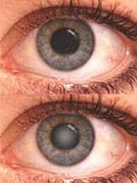
Its symptomatology is visual blurring, increase of the power of the lens with myopia or return of capability of close distance reading without glasses, reduced perception of blue color.
Patients complain of difficulty in reading, recognizing faces, their vision worsens in bright light, they experience haloes and sometimes monocular diplopia.
Sometimes cataract increases the converging power of the lens making the patient myopic.
Congenital or children cataract may interfere with development of vision and should undergo early surgery.
http://www.ncbi.nlm.nih.gov/pmc/articles/PMC2546248/?tool=pubmed
Cataract is an elderly disease and in the majority of the cases is idiopathic; in the adult may be consequent to trauma, drug-induced, (corticosteroid) related to internal or systemic diseases (Diabetes, Fabry's disease, Galactosemia (galactosemic cataract), Homocystinuria, Hyperparathyroidism, Hypervitaminosis D, Hypothyroidism, Mucopolysaccharidoses, Wilson's disease), related to skin diseases (Atopic dermatitis, Basal-cell nevus syndrom, Ichthyosis, Pemphigus) .
Cataract can begin in paediatric age and be congenital, due to intra uterus infections (Herpes, Syphilis, Citomegalovirus), gene defects or metabolic disorders; it may be isolated, or part of a syndrome.
Bilateral congenital cataract recognises more frequently a genetic cause.
Transmission is autosomical dominant, less frequently X-linked.
Congenital cataract is more frequent in the developing countries.
There are various anatomical types of cataract: nuclear, cortical and posterior subcapsular types.
Nuclear cataract is a nuclear type, caused by exaggerated sclerosis of the nucleus.
Here is a complete classification of the different forms of cataract.
http://www.ncbi.nlm.nih.gov/pmc/articles/PMC2546248/?tool=pubmed
http://en.wikipedia.org/wiki/Cataract#Classification
In one word, crystalline lens may be considered an overall marker of global health of the whole body; an opacity should prompt a screening for causes.
EPIDEMIOLOGY
Age-related cataract is responsible for 48% of world blindness, which represents about 18 million people, according to the World Health Organization (WHO); is the leading cause of blindness in the world.
http://www.who.int/blindness/causes/en/
In many countries, surgical services are inadequate, and cataracts remain the leading cause of blindness. Cataract incidence and prevalence increases with increase of mean age of the population.
Cataract is an important cause of low vision in both developed and developing countries.
Even where surgical services are available, low vision associated with cataracts may still be prevalent, as a result of long waits for operations and barriers to surgical uptake, such as cost, lack of information and transportation problems.
In the United States, age-related lenticular changes have been reported in 42% of those between the ages of 52 and 64,
http://www.ncbi.nlm.nih.gov/pubmed/7395962
in 60% of those between the ages 65 and 74,
http://www.ncbi.nlm.nih.gov/pubmed/879158
and in 91% of those between the ages of 75 and 85.
STRUCTURE OF THE LENS
Lens has a biconvex shape, consisting of two faces and a equatorial edge, where the zonular fibers are inserted. It consists of a capsule, an epithelial layer and a mass of cristallin fibers. Lens capsule is made of a scleroprotein like collagen.
Epitelial layer consists of a single layer of cells, with a transparent cytoplasm.
Lens fibers are epithelial cells, thin and long, clear and transparent, rich in water. Between lens fibers there is a cementing substance.
Lens has neither nerves nor vessels.
http://www.ncbi.nlm.nih.gov/pubmed/9486018
The transparency of the lens depends on a unique arrangement of tightly packed fibers wich rely on a certain protein structure; the lens is isolated in its environment by a capsule and epiteliuum. The damage of both structures(capsule, epitelium, fibers) may lead to the formation of cataract; the damage may be cumulative over the years.(H Cheng 1989)
The initiating mechanism of cataract may be different; end stages osmotic changes due to an influx of sodium are apparent in all cases. Lens volume depends on a balance of two forces: one is the permeability and the other is the active pump that extrudes sodium and concentrates potassium ions.
Experimental Ouabain treatment inhibites Na-K ATPase, blocking the pump mechanism and leading to lens swelling.
Also sugars, like glucose, galactose and xylose are cataractogenic; they enter the lens and are converted in alcohol that is no more able to exit the lens; hypertonicity is corrected by an influx of water.(Kinoshita 1974)
PATHOGENESIS
Cause of cataract is a loss of transparency; in aging eye epitelial cells density decrease leading to aberrant fiber formation.
Increasing age is the most important risk factor for age related cataract, as the opacity results from the cumulative damage of environmental insult to proteins and cells of the lens.
One common feature in all types of cataract is a dramatic change in hydration, due to osmotic changes for the influx of sodium and chloride ions.
In normal condition there is a balance between opposite forces; the permeability of the membrane and the cation pump.
Several factors can promote the formation of cataracts, including long-term exposure to ultraviolet light; all these mechanisms can be reconducted to inflammatory changes, oxidative damage, osmotic stress.
Photochemical insults, producing hydrogen peroxide superoxide, and hydroxyl radical induces damage to the lens epithelial cell DNA thus triggering a sequence of events leading to cataract.
Other mechanisms involved in cataractogenesis are increase of oxidation product and the conversion of soluble proteins into insoluble high molecular weight aggregates.
http://emedicine.medscape.com/article/1210914-overview#a0104
In experimental studies an inhibition of the DNA repair mechanisms of the lens has been observed (Santana 2011).
Elevated markers of systemic inflammation increase the risk of age-related cataract in otherwise health individuals. (Schaumberg 1999).
High serum levels of cytokine interleukin 6 predispose for age related nuclear cataract. (Klein 2006)
http://www.ncbi.nlm.nih.gov/pubmed/21779674
Furthermore, as the lens ages, a reduction in the rate at which water and, perhaps, water-soluble low-molecular weight metabolites can enter the cells of the lens nucleus via the epithelium and cortex occurs.
ETIOLOGY AND RISK FACTORS
http://www.ncbi.nlm.nih.gov.offcampus.dam.unito.it/pubmed?term=linda%20m%20meyer%2C%0Stefan%20%20lofgren%2C%20franz%20G%20Holz
Each cataract type has its own risk factors: nuclear cataract is associated with cigarette smoking; cortical cataract with UV exposure; posterior subcapsular with hypertension and steroid use (Klein 2012).
CONGENITAL CATARACTS
Different genetic patterns are associated with different types of cataract.(Klein 2012)
http://jama.jamanetwork.com/article.aspx?articleid=203039
Genetic factors are often a cause of congenital cataracts, that are the leading cause of reversible blindness in childhood; congenital cataract is visible at birth or in the first decade of life; may be isolated or associated with other ocular development anomalies or be part of a multisystem syndrome like Down syndrome or Wilson disease.
(http://www.ncbi.nlm.nih.gov/pubmed/21779674);
Positive family history may also play a role in predisposing someone to cataracts at an earlier age, a phenomenon of "anticipation" in presenile cataracts.
Among congenital cataracts an etiology was found in 62,5% of cases. Hereditary was the cause of 42,3% of etiologies. 77,7% of cases were autosomal recessive. 16,4% were associated with general diseases or dysmorphology syndromes. Metabolic diseases and intrauterine infections were 7 and 4,7% of cases. (El Fikh L, et al 2007)
Cataract is frequent in galactokinase deficiency childs, and prevented by a galactose-free diet.
http://www.ncbi.nlm.nih.gov/pubmed/5129188

May be a secondary effects of diseases such as diabetes, (http://www.ncbi.nlm.nih.gov/pubmed/22654491)
hypertension and advanced age; they are usually a result of denaturation of lens protein.
Cataracts may also be produced by eye surgical or traumatic injury (possibly much earlier).
Cataract may be caused by ionizing radiations, exposure to ionizing radiation, in case of X ray diagnostic exposure.
http://www.ncbi.nlm.nih.gov/pubmed/16971627 ;
Cataract was found in 335 of patients receiving radiotherapy for orbital lymphoma (Cohnen M, 2000), microwave, ultraviolet radiation like sunlight radiation; this explains the high prevalence of cataract in countries with hot climates (Cheng, 1989).
UVB-R may lead to cataract via protein modification, lipid peroxidation, DNA fragmentation induced by photooxidation.
Ultraviolet radiation may lead to formation of cromophores from proteins containing triptophan; the precise mechanism is still disputed, but may depend on the formation of free radicals scavengers setting in train reactions leading to protein aggregation. (Cheng, 1989)
UVB-R induced cataract may be caused by inflamamtory response, mediated by 1L-1 and IL-6 increased serum levels in experimental animals; exposition of one eye may cause on the same basis, cataract in the unexposed eye.
A study among Icelandair pilots showed commercial airline pilots are three times more likely to develop cataracts than people with nonflying jobs.
This is thought to be caused by excessive exposure at high altitudes to radiation coming from outer space, which becomes attenuated by atmospheric absorption at ground level.
A recent study on watermen in Chesapeake Bay shows correlation between cataract and exposition to sunlight (Hollows, lancet 1981); UV-A radiation was found fairly unimportant in cataract patogenesis.
http://www.ncbi.nlm.nih.gov/pubmed/16087845
Supporting this theory is the report that 33 of the 36 Apollo astronauts involved in the nine Apollo missions to leave Earth orbit have developed early stage cataracts that have been shown to be caused by exposure to cosmic rays during their trips.
At least 39 former astronauts have developed cataracts, of whom 36 were involved in high-radiation missions such as the Apollo missions.
http://science.nasa.gov/science-news/science-at-nasa/2004/22oct_cataracts/
Cataracts are also unusually common in persons exposed to infrared radiation, such as glassblowers, who suffer from exfoliation syndrome. Exposure to microwave radiation can cause cataracts. Atopic or allergic conditions are also known to quicken the progression of cataracts, especially in children. http://www.ncbi.nlm.nih.gov/pubmed/12546060
Cataract can also be caused by iodine deficiency.
http://www.ncbi.nlm.nih.gov/pubmed/1803312
Some drugs can induce cataract development, such as corticosteroids http://archopht.jamanetwork.com/article.aspx?volume=74&page=38
and the antipsychotic drug quetiapine (sold as Seroquel, Ketipinor, or Quepin).
Also smoking has been considered as a risk factor for cataract (Cheng 2000), specially pipe smoking, due to the larger amount of smoke produced by pipe-smokers; mechanism may be a direct toxic action on the lens by combustion of products like cadmium or hysocyanate, or by the continuous raise of temperature of the lens, or by an oxidative stress to the lens, reducing the levels of antioxidative components like ascorbic acid and nicotinamide.
http://www.hkmj.org/abstracts/v6n2/195.htm
This is probed by several case controlled, cross sectional and prospective studies. There is a dose-response between smoking and cataract.
http://www.ncbi.nlm.nih.gov/pmc/articles/PMC1836680/?tool=pubmed
http://www.sciencedirect.com.offcampus.dam.unito.it/science/article/pii/S0161642099900039
Inherited diseases: Steinert disease is the most frequent adult onsset muscolar distrophy (Prev 1/20000) and is characterized by myotonia and multiorgan damage i.e. muscle weakness, arrhythmias and cardiac disorders, cataract and endocrine disorders.
http://www.sciencedirect.com.offcampus.dam.unito.it/science/article/pii/S0755498207000036 http://www.ncbi.nlm.nih.gov.offcampus.dam.unito.it/pubmed??
Bilateral cataract is constant in fully developed disease, and may be the first and/or only manifestation of the disease.
It is a posterior subcapsular cataract, manifesting with multicolour opacities, increasing in number and size.
In Steinert disease DMPK gene expression is decreased, caused by the sequestration of messenger RNA into the cells with accumulation of proteins with citotoxic effect .
DMPK protein is located in cardiac muscle, in muscle cells, and into the lens.
http://www.ncbi.nlm.nih.gov/pubmed/17289339)
CATARACT AND METABOLISM
The most studied cataracts are those caused by high concentration of various sugars. The best known form of cataract occours in galactosemia wich results from a lack in enzyme galactose-1-phosphate urydiltransferase or galactokinase.
Galactokinase phosphorylates galactose to galactose-1-phosphate, which in turn can be converted to UDP-galactose, isomerized to UDP-glucose, incorporated in glycolipid or glycoproteins.
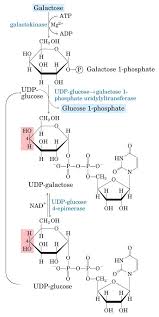
Galactosemia is an autosomal recessive disorder, characterized by hypergalactosemia and primary cataract formation at birth or in the first few months of life.
Cataract may be prevented by a galactose-free diet.
Most of the galactose that accumulates is converted to galactiol in the presence of aldose reductase wich accumulates in the lens and leads to increased osmotic pressure, hydration of the lens, as galactiol do not diffuse out of the capsule, and hyperosmotic disruption of the fibers.
An aldose reductase inibitor would prevent and reverse early cataract when instilled topically into the eyes of galactosemic rats.
Heterozygous form of galactokinase deficiency has been reported increased in Caucasian people developing idiopatic cataract between 20 and 55 years (Maraini 2003).
Diabetes mellitus complications involves every part of the eye; the most important complications are diabetic retinopathy and cataract.
Diabetes may affect lens transparency, due to diabetes itself or to an accelerated senile cataract. (Adeoti 2012); in diabetic patients cataract occurs 20 years earlier than in health population.
In diabetic patients under 60 Y cataract occurs three or four times that in the normal population (Cheng 1989); proposed mechanisms are overproduction of free radicals, decreased antioxidant capacity and oxidative damage by polyol pathway and advanced glycation end products. (Hashim 2012)

In diabetes oxidative stress plays a role in the pathogenesis of long term complications. Chronic exposure to glucose increases metabolic flux inducing mithocondrial suoperoxide production , subsequent damage to electron transport chain and accumulation of glycolitic intermediates.
Excessive production of reactive oxygen species produces oxidative stress increasing hexosamina and polyol pathway, formation of advanced glycation end products and glycation of proteins.
Polyol pathway is involved in cataractogenesis. Polyol pathway enzymes and aldose reductase and sorbitol dehydrogenase transform glucose to sorbitol and to fructose .
In normoglycemia 5% of glucose enters the polyol pathway where in hyperglycemia increases to 30%.
Sorbitol increases membrane permeability leading to cell lesions and eventually cataract (Hashim 2012)
Apart from osmotic damage, oxidative stress caused by polyol pathway play also a role in diabetes cataractogenesis.
Overactivity of aldose reductase depletes the cofactor NADPH, necessary for the regeneration of reduced glutathione by glutathione reductase cellular antioxidant capacity is reduced.
These metabolic changes have been found in diabetic patients lenses.
Moreover glycation of lens proteins leads to aggregation, cross linking and insolubilization.
Diabetes is a well estabilished cause of cataract in most clinic based and population based studies (Leske 1998).
http://www.sciencedirect.com.offcampus.dam.unito.it/science/article/pii/S0161642099900039
Diabetes is associated with overall lens changes, specially with cortical and posterior subcapsular opacities; is not clear the relation with nuclear opacity.
In cataract some studies recognized a role to magnesium.
In age related cataract a ionic imbalance has been shown, with decreased magnesium and potassium and increased calcium and sodium, due to membrane changes and leading to lens opacification.
As already seen, high amount of free radicals are generated in lens due to UVB exposure, but antioxidative mechanisms may prevent the damage.
Excessive production of free radicals, as H2O2, nitric oxide (NO), and reduced antioxidant capacity like superoxide dismutase, catalase, glutathione predispose to oxidative damage.
Magnesium deficiency may trigger the cataractogenesis increasing the iNOS expression and release of NO.
Magnesiun deficiency affects activity involved in ATP synthesis, reducing ATP levels and the activity of the Na+/K+ ATPase. Sodium accumulates intracellularly, causing osmotic stress.
http://www.ncbi.nlm.nih.gov/pubmed/17289339
PROGNOSIS AND COMPLICATIONS
If not treated, cataract leads to complete blindness.
THERAPY
Medical therapy finds little space in cataract therapy
Topical treatment (eye drops) with the less well-known antioxidant N-acetylcarnosine has been shown in randomized controlled clinical trials to lens transparency for patients with cataracts. http://www.ncbi.nlm.nih.gov/pubmed/11390029
After animal experiments researchers of a pharmaceutical company have proposed N-acetylcarnosine as a treatment for ocular disorders that have a component of oxidative stress in their genesis, including cataracts, glaucoma, retinal degeneration, corneal disorders, and ocular inflammation.
http://www.ncbi.nlm.nih.gov/pubmed/10851037
Statin have been shown to have antioxidant activity; nitric oxide derived oxidant species that are active in atherogenesis are suppressed by statins. Oxidative stress is thought to be related to age related nuclear cataract experimental studies demonstrate inverse correlation between statin use and incidence of nuclear cataract; .
Statin use do not affect cortical or posterior subcapsular cataract. (Klein 2012) http://jama.jamanetwork.com/article.aspx?articleid=203039 ;
Nowadays, surgery is the only therapy for cataract; surgery began in 1960 when Svyatoslav Fyodorov performed the first intraocular lens replacement operation.
The operation to remove cataracts can be performed at any stage of their development. There is no longer a reason to wait until a cataract is "ripe" before removing it.
http://www.rnib.org.uk/eyehealth/eyeconditions/conditionsac/Pages/cataract.aspx
because the old concept of “ripe” cataract is no longer valuable and surgery should be tailored on the individual patient (Elkington 1998)
Two types of eye surgery can be used to remove cataracts: extracapsular cataract extraction (ECCE) and intracapsular cataract extraction (ICCE).
ECCE surgery consists of removing the lens, but leaving the majority of the lens capsule intact. High frequency sound waves (phacoemulsification) are sometimes used to break up the lens before extraction.
Intra-capsular (ICCE) surgery involves removing the lens and lens capsule, but it is rarely performed in modern practice.
In either extracapsular surgery or intracapsular surgery, the cataractous lens is removed and replaced with a plastic lens an intraocular lens implant.
Cataract operations are usually performed using a local anaesthetic, and the patient is allowed to go home the same day.
Complications are possible after cataract surgery, including endophthalmitis, posterior capsular opacification and retinal detachment
Capsular secondary cataract is removed with a laser beam.
CURIOSITY
Cataract surgery: 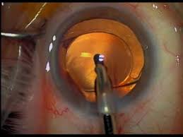
http://www.youtube.com/watch?v=evTf6Qse4rg
BIBLIOGRAPHY
1. A.R. Elkington, P.Khaw; ABC of eyes; cataracts Br Med J 1988; vol 296: 1787-1790
2. Agarwal R., Iezhitsa I, Agarwal P et al. Magnesium deficiency: does it have a role to play in cataractogenesis? Exp Eye Res 2012; jun 2 (in press)
3. Asdeoti C O, Isawumi M A, Ashaye AO et al. the anterior segment of the eye in diabetes. Clin Ophtal 2012; 6: 667-671Galactiokinase deficiency in a newborn infant. Arch Dis Childhood 1971; 46: 864
4. Bianchi C, Bandello F, Brancato R Manuale di oftalmologia, Ghedini editore, Milano, 1995
5. Bouhour F, Bost M, vial C. Maladie de Steinert. La presse medicale 2007 : 36 n 6 : 965-971
6. Branka S.K. Ocular abnormalities and systemic diseases in Down sindrome Sindrome Strabismus 2012; 20(2): 74-77
7. Cheng A C K, Pang C P, Leung A T s et al: The association between cigarette smoking and ocular diseases. H K M J 200; 6 n 2: 195-202
8. David A. Quillen , M.D.: Common Causes of Vision Loss in Elderly Patients; Am Fam Physician. 1999 Jul 1;60(1):99-108.
9. Duncan G, Wormstone M, Davies P D. The aging human lens: structure, growth and physiological behaviour. Br J Ophtal 1997; 81: 818-23
10. Ederer F, Hiller R. Senile elns changes and diabetes in two population studies. Am J ophtal 1981; 91(3): 381-95Romeo V Myotonic dystrophy type 1 or Steinert disease. Adv Exp Med boil 2012; 724: 239-57
11. El Fkih L, Hmaied W, Moalla S, et al. Congenital cataract etiology. Tunis Med 2012; 85(12): 1025-Cohnen M, Wittsack HJ, Assadi S. rsadiation exposure of patients in comprehensive computed tomography of the head in acute stroke. AJNR 2006; 27: 1741-45Santana A, the genetic and molecular basis of congenital cataract. Arq Bras oftalmol 2011; 74(21): 136-42
12. Elkington A R, Khaw P T ABC of eyes: cataracts. Br Med J 1988; 296. 1787-1790
13. H Cheng Causes of cataract. BMJ 1989; 298: 1470-71
14. Hashim Z, Zarina S. Osmotic stress induced oxidative damage: possible machenism of cataract formation in diabetes. J diabetes compl 2012; May 17 (in press)
15. Hashim Z, Zarina S. Osmotic stress induced oxidative damage: possible mechanism, of cataract formation in diabetes. J Diab and its complications 2012; (in press)Hodge W G, Whitcher J P, Satariano W. Risk factors for age related cataracts. Epidemiol Rev 1995; 17(2): 336-46
16. Kaushik M et al. Risk of radiation retinopathy in patiernts with orbital and ocular lymphoma Int J radiat oncol biol phis 2012; 5: in print
17. Kinoshita JH. Mechanisms initiating cataract formation. Proctor Lecture. Invest Ophthalmol. 1974 Oct;13(10):713-24
18. Klein B, Klein R, Lee K E et al. Statin use and incident nuclear cataracrt. JAMA 2006; 295: 2752-2758
19. Klein BE, klein R, Lee K et al. Markers of inflammation, vascular endotelial dysfunction and age related cataract. AJOphtalmol 2006; 141: 116-122
20. Leske M C, Suh Yuh Wu M A, Anselm H et al. Diabetes, hypertension and central obesity as cataract risk factors in a black population. Ophtalmology 1999; 106: 35-41
21. Maraini G, Fielding Hejtmancik J, Shields A et al. Galactokinase gene mutation and age related cataract. Lack of correlation in an italian population. Molecular vision 2003; 9: 397-400
22. Meyer L et al. Bilateral cataract induced by UVR-B exposure –evidence for inflammatory response. Acta ophtalmol mar 2012 (in press)
23. Schaumberg D, Ridker P, Glynn R et al. High levels of plasma C-reactive protein and furture risk of age-related cataract. Ann Epidemiol; 1999.; 9:166-171
statin use: http://www.ncbi.nlm.nih.gov.offcampus.dam.unito.it/pubmed/16788130