DEFINITION
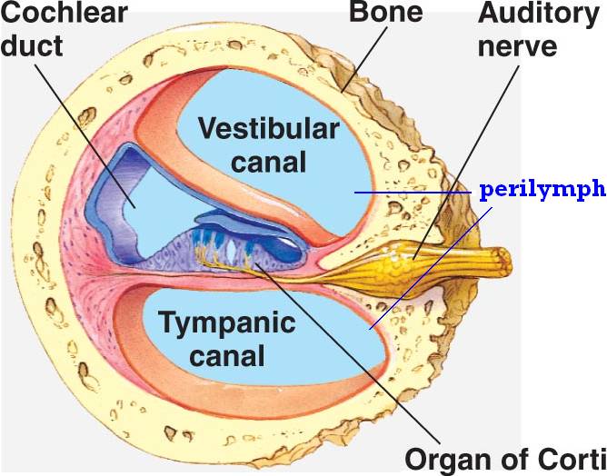
The Organ of Corti is a part of the cochlea and it mediates the sense of hearing transducing pressure waves to action potentials. This structure is localized in the scala media and it is formed by a series of hair cells, nervous terminations of spiral ganglion and supporting cells.
GROSS ANATOMY
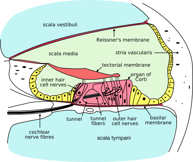
The scala media, or cochlear duct, is located between scala tympani and scala vestibuli and it is filled with endolymph. This structure is delimited by the basilar membrane and Reissner’s membrane. The Organ of Corti covers the basilar membrane and it is under the tectorial membrane, an acellular gel into which hair cell stereocilia are immersed. The peripheral process of acoustic nerve fibers provides synaptic connections between hair cells and cochlear nucleus.
The upper portion of the cochlear duct is formed by the stria vascularis, which contains numerous capillary loops and small blood vessels and produces endolymph.
Frequency-dependent properties of the tectorial membrane facilitate energy transmission and amplification in the cochlea.2013
HISTOLOGY
Organ of Corti consists of different types of cells:
*Inner hair cells
*Outer hair cells
*Supporting cells

Inner Hair Cell
These cells are specialized in the mechanoelectrical transduction. There are almost 3500 cells disposed in one line along all the basilar membrane. They are connected to type I neuron peripheral fibers of spiral ganglion, these connection are very divergent (10/1). The luminal part of the cell is immerged in endolymph, the basal one is immerged in normal extracellular fluid. The luminal portion is formed by bundles of stereocilia(inner_ear), whose tips are connected by filamentous structures called tip-links.

Molecular remodeling of tip links underlies mechanosensory regeneration in auditory hair cells.2013
The composition and role of cross links in mechanoelectrical transduction in vertebrate sensory hair cells.2013
Outer Hair Cell
These cells are acoustical pre-amplifiers. They are almost 12000, disposed in three parallel lines. These cells are connected to type II amyelinic neurons, the connections are very convergent. They have also an afference from superior olivary nucleus. They have contractile activity.

Myosin light chain kinase regulates hearing in mice by influencing the F-actin cytoskeleton of outer hair cells and cochleae.2014
Supporting Cells
These cells are of four different types: Corti pillars, Hensen cells, Deiters cells and Claudius cells.
Endolymph
Endolymph fills the scala media and it is produced by stria vascularis.
Potassium secreted into the endolymph by the stria vascularis enters the hair cells through apical mechanosensitive channels. It is recycled back to the stria vascularis through supporting cells and fibrocytes of the spiral ligament for another round of secretion. Hair cells and stria vascularis are tied together in a "push-pull" or "pump-leak" balance that determines endocochlear potential (EP +85mV), endolymph composition and ultimately the sensivity and stability of hair cells and hearing over a lifetime.
The evolutionary strategy of using K receptors currents is due to the fact that it reduces metabolic requirements because it has a passive outflow from hair cells to the basal membrane, instead of active Na extrusion.
Endolymph has a particular ion concentration
| Ion | Perilymph (mM) | Endolymph (mM) |
| Na | 154 | 1 |
| K | 3 | 161 |
| Cl | 128 | 131 |
There is a potential difference of -140 mV between endolymph and inner receptor.

A basolateral Na/K-ATPase pumps K and creates a Na gradient to drive K and Cl into the cell in a co-transport process. Chloride is recycled by basolateral Cl channels, and K is secreted apically by KCNQ1/KCNE1 K channels, which are open at the unusual apical membrane voltage of marginal cells (inside positive by about +10 mV). This allows for a net secretion into the endolymph in spite of its high K concentration.
KCNQ proteins have six transmembrane domains and a pore-forming P-loop. Mutations on KCNQ1 and KCNQ4 lead to deafness. Indeed KCNQ4 was mapped to human chromosome 1p34; it is one of over 30 loci for dominant deafness.
Hypoxia reduces endocochlear potential, due to a drop in metabolism and strial current; whereas a depletion of vascular K does not drop EP, showing that K comes from spiral ligament rather than from blood.
Neuronal KCNQ potassium channels: physiology and role in disease. 2000
Ion flow in stria vascularis and the production and regulation of cochlear endolymph and the endolymphatic potential.2011
SENSE OF HEARING
The role of the sense of hearing is translating pressure waves of perilymph and endolymph to electrical signal and acoustic sensation.

Mechanism of transduction

- Deflection: tip-links open K channels and k enters the cell
- Depolarization: Ca enters the cell through voltage-dependet channels
- Higher Ca concentration: K extrusion toward basolateral portion and Glutamate release in synaptic cleft
- Action potential along acoustic nerve (VIII)
ABNORMALITIES
Deafness is a partial or total inability to hear.
AHL is the age-related hearing loss, also known as presbycusis. Many K channels, like KCNQ1, were found to be down-regulated in stria vascularis with aging. Moreover a reduced KCNQ4 current may lead to a slow degenerative process. Although in some cases the EP is maintained and this is a paradoxical phenomenon, it would appear that this is attributable to the SV which may generate a much lower current to establish a new balance for potassium influx and efflux at a relatively lower level. It is not clear if an atrophied stria vascularis could recover sufficient function by up-regulating these potassium transporters expression to maintain a normal endocochlear potential.
See also Deafness and Apoptosis
Prestin regulation and function in residual outer hair cells after noise-induced hearing loss.2013
Compromised potassium recycling in the cochlea contributes to conservation of endocochlear potential in a mouse model of age-related hearing loss.2013
EMBRIOLOGY
Otic vescicle
Auditory ganglion source of Sonic hedgehog regulates timing of cell cycle exit and differentiation of mammalian cochlear hair cells.2013