Serum Uric Acid
Blood Tests

Author: Gianpiero Pescarmona
Date: 11/10/2009

Description

DEFINITION

A short yet comprehensive description

External links

DatabaseLink
"XYZZ":"test":
"ZXVX":"URL":

ANALYTICAL METHOD

ANALYTICAL TRICKS AND TIPS

THE BIOLOGICAL CONTEXT
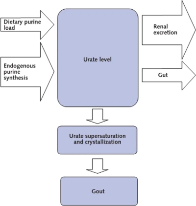
Gout Development

THE NORMAL RANGE

DIAGNOSTIC USE

Issues

Specificity, sensitivity etc.

Diagnostic Algorithms

PROs and CONTROs

Open Questions

Association between vitamin D insufficiency and elevated serum uric acid among middle-aged and elderly Chinese Han women. 2013

Vitamin D insufficiency was significantly associated with elevated uric acid among postmenopausal Chinese Han women.

Stabilita associazione tra livelli di acido urico e diabete
Livelli sierici elevati di acido urico risulterebbero associati allo sviluppo di diabete di tipo 2. Sono i risultati di un'ampia metanalisi pubblicata da un gruppo di ricercatori giapponesi su Diabetes Care. L'indagine ha preso in considerazione ben 11 studi clinici, presenti nei database Medline (dal 1966 al 2009) ed Embase (dal 1980 al 2009), riguardanti oltre 42mila pazienti diabetici e con follow-up compresi tra 2,0 e 13,5 anni. In sintesi, gli autori hanno calcolato un rischio relativo di sviluppare diabete pari a 1,17 per ogni incremento di 1 mg/dL di acido urico nel siero. "Trattandosi di studi osservazionali non è stato possibile stabilire una relazione causa-effetto tra livelli di acido urico e diabete" ha commentato Hirohito Sone del Department of Internal Medicine, University of Tsukuba di Ibaraki. "Per stabilire se i livelli di questa sostanza possano rappresentare un fattore predittivo dello sviluppo di diabete sarà necessario valutare non solo la suddetta relazione causa-effetto ma anche i valori di cut-off di acido urico". (L.A.).

Diabetes Care 2009, 32, 1737-1742

Serum uric acid is associated with bone health in older men: A cross-sectional population-based study. 2011 J Bone Miner Res. 2011 May;26(5):955-64. doi: 10.1002/jbmr.286.

Serum uric acid (UA) is a strong endogenous antioxidant. Since oxidative stress has been linked to osteoporosis, we examined the association between serum UA levels and bone mineral density (BMD), prevalent vertebral and nonvertebral fractures, and laboratory measures such as calcitropic hormones and bone turnover marker levels. This cross-sectional analysis consisted of 1705 community-dwelling men aged 70 years or over who participated in the baseline part of the Concord Health and Ageing in Men Project (CHAMP), a population-based study of older men in Sydney, Australia. BMD at all sites was significantly higher among men with serum UA levels above the group median than among men with UA levels below the median. In multiple regression analyses adjusted for potential confounders, serum UA remained associated with BMD at all sites (β = 0.12 to 0.14, p < .001), serum calcium (β = 0.11, p = .001), parathyroid hormone (β = 0.09, p = .002), 25-hydroxyvitamin D (β = 0.09, p = .005), and was negatively associated with urinary excretion amino-terminal cross-linked telopeptide of type 1 collagen (β = -0.09, p = .006). Overall, serum UA accounted for 1.0% to 1.44% of the variances in BMD (R(2)  = 0.10 to 0.22). In multiple logistic regression analyses, above-median serum UA levels were associated with a lower prevalence of osteoporosis at the femoral neck [odds ratio (OR) = 0.42, 95% confidence interval (CI) 0.22-0.81, p = .010) and lumbar spine (OR = 0.44, 95% CI 0.23-0.86, p = .016) and a lower prevalence of vertebral (OR = 0.62, 95% CI 0.43-0.91, p = .015) and nonvertebral (OR = 0.51, 95% CI 0.29-0.89, p = .018) fractures. In conclusion, higher serum UA levels are associated with higher BMD at all skeletal sites and with a lower prevalence of vertebral and nonvertebral fractures in older men. © 2011 American Society for Bone and Mineral Research.

Working Hypothesis

Metabolite profiling reveals new insights into the regulation of serum urate in humans, 2014

Comments
2014-06-09T13:16:16 - Elena Rubini

DEFINITION
Uric acid , a weak organic acid with a pka of 5.75, is the end product of purine degradation. It is a heterocyclic compound of carbon, nitrogen, oxygen and hydrogen, with the formula C5H4N4O3.

Fig.1 Uric Acid molecule

RANGE
Its plasma concentration is defined and uric acid in the human being is normally between 2.5 and 7.0 mg / dl in men and between 1.5 and 6.0 mg / dl in females. The limit of 7.0 mg / dl is important, because at that concentration the saturation occurs and precipitation of crystals typical of gout becomes possible.
In case of higher values we speak about hyperuricemia; in case of lower levels about hypouricemia.
Hyperuricemia is a common finding in patients with the metabolic syndrome and an inverse correlation was noted between insulin resistance and decreased renal uric acid clearance, which is itself associated with elevated uricemia.
Obesity, in particular visceral adiposity, is also positively associated with hyperuricemia, which can be reduced by body weight loss. Hyperuricemia is also frequently observed in patients with cardiovascular diseases.

SYNTHESIS
Uric acid is a breakdown product of purines and uric acid generation therefore depends directly on both intrinsic purine production and purine intake. In humans, uric acid is an end-product metabolite; consequently the depletion of uric acid depends directly on its excretion. The balance between uric acid production and excretion determines the serum urate level.

Purines are heterocyclic aromatic compounds, consisting of conjoined pyrimidine and imidazole rings. In mammals, the most common expression of purines is found in the form of deoxyribonucleic acid (DNA) and ribonucleic acid (RNA) (containing the purines adenine and guanine).
Purines are also critical elements of the energy metabolism molecules NADH, NADPH, and coenzyme Q, and it may also serve as direct neurotransmitters; for example, adenosine may interact with receptors to modulate cardiovascular and central nervous system function.

Fig.2 Purines examples

Purine biosynthesis is initiated on a core of ribose-5-phosphate. The enzyme phosphoribosyl pyrophosphate (PRPP) synthase catalyzes the addition of a pyrophosphate moiety to form the adduct PRPP. Subsequently, the enzyme glutamine-PRPP amidotransferase catalyzes the interaction of PRPP with glutamine to form 5-phosphoribosyl amine, next it forms the backbone for a series of molecular additions, ending in the formation of the purine inosine monophosphate (IMP). IMP is converted into either adenosine monophosphate (AMP) or guanosine monophosphate (GMP), which can then be phosphorylated into higher-energy compounds.

Fig.3 Purines biosynthesis

Purines are susceptible to enzymatic catabolism, presumably to maintain purine homeostasis:
GMP and IMP are converted by nucleotidases to their purine base forms, guanosine and inosine. Instead AMP is not susceptible to nucleotidase activity, but it can undergo conversion, through the activity of adenylate deaminase, into IMP for further degradation. Additionally, adenosine deaminase can convert adenosine to inosine for inclusion in the degradative pathway. Further catabolism of both guanosine and inosine is mediated by the common enzyme purine nucleoside phosphorylase. Guanosine is converted to guanine, whereas inosine is converted to hypoxanthine. Both guanine and hypoxanthine are subsequently converted to xanthine, by the enzymes guanine deaminase and xanthine oxidase, respectively. Xanthine from either source is then converted directly to uric acid, again by the action of xanthine oxidase.

Fig.4 Purines salvage

Purine biosynthesis not only directly increases the substrate load for urate generation but also increases the turnover of already-formed purines that contribute to increased urate levels.

TRANSPORT AND EXCRETION
Urate homeostasis really depends on the balance between production and complex processes of secretion and reabsorption in the kidney tubule and excretion in the intestine. It is estimated that approximately 30% of uric acid excretion is by the intestine, renal mechanisms of urate excretion account for the other 70%.
The gastrointestinal action may represent a minor pathway for urate excretion under most circumstances, it may become more important in settings of renal insufficiency. Mechanisms of uric acid transport into the gut appear to include exocrine secretion (saliva, gastric, and pancreatic juices), as well as direct bowel secretory mechanisms. Uric acid is apparently excreted into the gut in its native form and then undergoes degradation by intestinal flora.

Urate handling involves urate glomerular filtration followed by a complex array of reabsorptive and secretory mechanisms taking place in the proximal tubule.
In the bloodstream, uric acid (in the form of urate anion) is considered to be completely or nearly completely unbound to plasma proteins. As a result, nearly 100% of the urate load entering the renal afferent arteriole undergoes ultrafiltration by the glomerulus, next urate undergoes several distinct handling steps:

(1) a resorption step, in which as much as 90% to 98% of the filtered urate undergoes reclamation
(2) a secretion step, in which most of the urate resorbed in step 1 is retransferred back into the tubule lumen
(3) a possible additional resorption step in which a smaller amount of uric acid is then resorbed. The net result is excretion of approximately 10% of the filtered load.

The urate reabsorption pathway involves the apical exchanger proteins URAT1, OAT4, and OAT10: URAT1 is a 12-transmembrane domain–containing protein found in the apical membrane of proximal tubule epithelial cells and transports urate in exchange for Cl– or organic anions. Urate uptake by URAT1 and OAT10 is accelerated by intracellular monocarboxylates such as lactate, pyrazinoate, and nicotinate.
OAT4 is a multispecific anion transporter present in the apical membrane of epithelial cells from the proximal tubule. It is involved in luminal urate reabsorption by a mechanism that is transactivated by intracellular dicarboxylates but not by the antiuricosuric agent.
Urate transport by URAT1, OAT4, and OAT10 would lead to accumulation of urate intracellularly and presumably to gradients that would eventually impair further urate uptake if a mechanism did not exist to transport intracellular urate out of the cell at the basolateral surface.

This function appears to be served by Glut9a, which was first identified as a glucose transport family protein, but has little or no glucose transport capacity. Instead, Glut9a is an effective transporter of urate from the renal epithelial cell out into the renal interstitium.
GLUT9 is also involved in the transport of fructose, It is possible that this finding may contribute to understand the associations of hyperuricemia, gout and components of metabolic syndrome in diets rich in fructose, epidemiologic studies confirm a role for fructose consumption in hyperuricemia; patients who consume excessive fructose in the form of fructose-sweetened soft drinks or fruit juices demonstrate both higher serum urate levels and increased incidence of gout.
GLUT9 exists as two alternatively spliced variants that encode different aminoterminal cytoplasmic tails, but GLUT9a is expressed in the basolateral membrane, whereas GLUT9b is targeted to the apical pole.

A significant relationship was found between increased expression of the GLUT9b, but not GLUT9a, isoform and plasma uric acid levels.

In humans, GLUT9b expression is restricted to liver and kidney, whereas GLUT9a has a broad tissue distribution including liver, kidney, intestine, leukocytes, and chondrocytes, where its expression is upregulated by inflammatory cytokines 

Several apical monocarboxylate transporters are required to favor urate reabsorption, such as MCT9 and SMCT1 and 2.

Other transport proteins in the renal tubule epithelium regulate the excretion of urate from the peritubular fluid into the tubular lumen. At the basolateral surface, OAT1 and OAT3 transport urate from the interstitium into the epithelial cell cytosol. These transporters act via exchange with dicarboxylate anions and transport not only urate, but other organic anions and some drugs.

The multidrug resistance protein MRP4, a member of the superfamily of ATP-binding cassette (ABC) transporters, mediates ATP-dependent urate transport. ABCG2, a urate efflux transporter, which is expressed in the apical membrane of proximal collecting duct cells, also directly mediates urate excretion. Genetic association studies have implicated two other anion transport proteins as playing a role in apical urate transport outside of the cell, namely, NPT1 and NPT4. Additionally, genetic studies have implicated nontransporter proteins as playing roles in urate excretion including PDZK1, CARMIL, NHERF1, SLC5A8 and SLC2A12. The association of PDZK1, NHERF1, CARMIL, and the urate transporters has been suggested to form an apical transportasome complex implicated in the regulation of urate transport . This complex also includes the sodium monocarboxylate cotransporters (SMCT1, SLC5A8 and SMCT2, SLC2A12).

These genetic and biochemical data thus indicate that very complex regulatory processes may control the magnitude and direction of urate fluxes across the proximal tubule epithelium.

Renal transport of uric acid

Attachments
fileuserdate
GTR9_1_e_2_clustalw.pnggp15/06/2014
AddThis Social Bookmark Button