Definition
Pemphigoid is a group of uncommon and rare autoimmune blistering skin diseases. As its name indicates, pemphigoid is similar to pemphigus, but unlike pemphigus, pemphigoid does not feature acantholysis or degeneration of basal cells layer.

Epidemiology
Pemphigoid is less common than pemphigus, and is slightly more common in women than in men. The new cases are about 6-7/million/year. It is also more common in people over 60 years of age than it is in younger people.
Pathogenesis
Patients with BP have circulating IgG and IgE autoantibodies against 2 hemidesmosome proteins, BP230 (also known as BPAG1) and BP180.
BP230 is a plakin protein family member that promotes the association of hemidesmosomes with keratin intermediate filaments.
BP180 is a type II, transmembrane collagen that is associated with hemidesmosome–anchoring filament complexes. It has a long carboxy-terminal collagenous domain that projects into the extracellular region beneath the epidermal hemidesmosome. The collagenous domains have the characteristic tripeptide repeat, Gly-X-Y. A normal proteolytic processing event results in the shedding of the BP180 extracellular domain (LAD1) from the keratinocyte cell surface. The biologic relevance of this process is not yet known. The interactions of BP180 with other constituents of the anchoring complex have been extensively studied and underscore the importance of BP180 in the assembly and functioning of this cell-matrix adhesion structure. In addition to its role in maintaining the integrity of the dermal-epidermal junction, there is evidence that BP180 is involved in transmembrane signal transduction and in the regulation of keratinocyte differentiation. The extracellular domain of this protein contains 15 interrupted collagenous domains. Rotary shadowing studies of purified BP180 image its intracytoplasmic region as a globular head and its ectodomain as a central rod joined to a flexible tail. Immunoelectron microscopy studies indicate that BP180 spans the lamina lucida and inserts into the lamina densa.
BP180 is targeted by autoantibodies from patients with BP, pemphigoid gestationis, cicatricial pemphigoid, and linear IgA dermatosis.

Schematic overview of the plasminogen/plasmin activation cascade in experimental murine BP. Anti-BP180 IgG binds epidermal BM, activates complement, and generates neutrophil-rich infiltrates in skin. Subsequently, plasmin activates MMP-9, which in turn inactivates α1–proteinase inhibitor (α1-PI), thus allowing unrestrained activity of neutrophil elastase that degrades BP180 and produces subepidermal blisters. tPA, tissue plasminogen activator; uPA, urokinase plasminogen activator.
Epitope mapping studies of recombinant proteins have previously shown that autoantibodies from most patients with BP bind a determinant within the sixteenth noncollagenous domain of BP180 (i.e., the portion of its ectodomain that is positioned adjacent to plasma membranes of basal keratinocytes).

Autoreactive T and B Cells from Bullous Pemphigoid (BP) Patients Recognize Epitopes Clustered in Distinct Regions of BP180 and BP230, 2006
Passive transfer of experimental IgG developed against the murine homolog of this determinant to neonatal BALB/c mice produces clinical, histologic, and immunopathologic alterations like those seen in patients with BP. Antibody-induced blister formation in this animal model is dependent upon the activation of complement, degranulation of dermal mast cells, and generation of neutrophil-rich infiltrates.

Schematic model of the epidermal BM. The major subregions of epidermal BM are depicted in the context of autoimmune and genetic blistering diseases that develop as a consequence of acquired or inherited impairments in proteins within this cell-matrix adhesion junction. AECP, anti-epiligrin cicatricial pemphigoid; CP, cicatricial pemphigoid; EB, epidermolysis bullosa; IB, immunobullous; LAD, linear IgA dermatosis; OCP, ocular cicatricial pemphigoid. GABEB, generalized atrophic benign epidermolysis bullosa; PA, pyloric atresia. (J Clin Invest. 2005 April 1; 115(4): 825–828. doi: 10.1172/JCI200524855. Copyright © 2005, American Society for Clinical Investigation).
BP180
CHEMICAL STRUCTURE AND IMAGES
When relevant for the function
- Primary structure
- Secondary structure
- Tertiary structure
- Quaternary structure
Protein Aminoacids Percentage
The Protein Aminoacids Percentage gives useful information on the local environment and the metabolic status of the cell (starvation, lack of essential AA, hypoxia)
Model (Width 600 px)

mRNA synthesis
protein synthesis
post-translational modifications
degradation
CELLULAR FUNCTIONS
cellular localization,
biological function
- Enzymes
- Cell signaling and Ligand transport
- Structural proteins
REGULATION
http://www.ncbi.nlm.nih.gov/gene/1308
http://www.ncbi.nlm.nih.gov/pubmed/1324962


Molecular weight: 150,418.98
Clinical findings
Symptoms


Some people may have no symptoms, others may have mild redness and irration.
In severe cases, they are multiple blisters that may break open and form ulcers or open sores. Bullous is the medical term for a large blister (a thin-walled sac filled with clear fluid). Usually the skin in BP is very itchy and large, red welts and hives may appear before or during the formation of blisters.
 The blisters are widespread and usually appear on the areas of the body that flex or move (flexural areas), usually on the arms, legs or middle of the body About 15-20 percent of people with BP also develop blisters in the mouth or down the throat in the esophagus.
Other symptoms may include:
- Itching
- Rashes
- Mouth sores
- Bleeding gums
http://www.nlm.nih.gov/medlineplus/ency/article/000883.htm
Signs
D.D with Pemphigus because of negative Nikolsky sign (pressing a finger on the skin of the patient doesn't make the superficial layer move from the derma)
Associated conditions
Some patients with BP have other autoimmune diseases such diabetes and rheumatoid arthritis. Various other factors have been reported to play a role in triggering BP. These include drugs (furosemide, penicillin's), mechanical trauma, and physical traumas (burns from radiation, sun or heat).
Recent studies (Exp Dermatol. 2008 May;17(5):446-54 Searching for foreign antigens as possible triggering factors of autoimmunity: Torque Teno virus DNA prevalence is elevated in sera of patients with bullous pemphigoid.) report that the Torque Teno Virus (TTV) might be involved in the triggering of the skin-specific autoimmunity because of matching regions for the major BP antigens BP180 and BP230
Diagnosis
Because of all the variations and differing degrees of symptoms, the diagnosis must be confirmed by skin biopsy. A special skin biopsy test (a direct immunofluorescence biopsy) may also be needed. Blood tests are usually inconclusive.
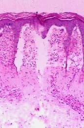
Biopsies of lesional skin show subepidermal blisters that are either granulocyte-rich or granulocyte-poor, depending on whether the biopsies were obtained from inflamed or noninflamed skin.
Direct immunofluorescence microscopy of perilesional skin shows linear deposits of IgG and/or complement component C3 in epidermal BM.
(http://www.ncbi.nlm.nih.gov/pmc/articles/PMC1070439/?tool=pmcentrez)
Treatment
The treatment of this disease includes:
- Corticosteroids
- Immunosuppressive drugs
- Plasmapheresis
- Appropriate skin care
Therapy of bullous diseases consists of suppressing the immune system, controlling inflammation and improving healing of erosions. The therapeutic agents used in the treatment of bullous diseases may be associated with high morbidity and occasional mortality. Successful treatment requires understanding of the pathophysiology of the disease process and the pharmacology of the drugs being used.
Bullous pemphigoid (BP) results from an autoimmune response and is mediated by a prominent cellular inflammatory infiltrate rich in eosinophils. Unlike PV, BP may remit spontaneously. Unlike therapy for PV, effective treatment of BP may be accomplished with lower doses of immunosuppressive drugs and/or anti-inflammatory agents. Potent topical steroids should be considered and favored in the management of localized or limited disease
Most patients with generalized BP require systemic therapy, the most commonly used agents being glucocorticoids (Church 1960; Siegel and Eaglstein 1984). Systemic glucocorticoids are usually sufficient as the sole therapy in the majority of cases. The dose is 0.5–0.75 mg/kg/day. Unlike patients with PV, higher doses of prednisone are rarely needed. A clinical response is usually attained rapidly (within 1–3 weeks) and is heralded by healing of existing lesions and cessation of new blister formation. The prednisone dose is then gradually tapered to 5mg daily over a period of 5–6 months
Immunosuppressive drugs are rarely needed in patients with BP. They may be used in patients who require high maintenance doses of glucocorticoids, for patients who are experiencing adverse effects from glucocorticoids, and for those whose disease does not respond completely to glucocorticoid therapy. The most commonly used agents are azathioprine (Greaves et al 1971; Burton and Greaves 1974), MMF (methylmicophenolate) (Nousari et al 1998; Wojnarowska et al 2002), and methotrexate (Downham and Chapel 1978)
MMF is effective both as a sole agent as well as in combination with glucocorticoids (Nousari et al 1998; Grundmann-Kollman et al 1999; Nousari, Brodsky, et al 1999; Nousari, Sragovich, et al 1999). The dose is usually 1 g twice daily. The dose of azathioprine is 1–3 mg/kg daily. The dose may be adjusted based on the clinical response, side effects and the level of TPMT. Methotrexate is effective in relatively small doses (up to 12.5 mg weekly) as a sole therapy (Heilborn et al 1999)
In general, once the patient who is receiving prednisone and an immunosuppressive agent attains complete remission, the prednisone is tapered and discontinued. The immunosuppressive agent is maintained and ultimately slowly tapered and discontinued.
Plasmapheresis may be used in severe cases and is usually used in association with immunosuppressive drugs (Roujeau et al 1984; Goldberg et al 1985). Plasmapheresis is costly, time consuming, and only temporarily beneficial.
Appropriate skin care is important in the healing of lesions and avoidance of complications. After blisters rupture, resulting erosions may become secondarily infected with bacteria, may progress to ulcers (especially over pressure areas such as the buttocks and the back), and may acquire crusting that interferes with reepithelialization and healing. Measures that help in the healing process include gentle cleansing of the erosions, liberal application of antibiotic ointments or petroleum jelly, and avoidance of trauma. The elderly are especially prone to complications and may require hospital admission for proper skin care.