

DEFINITION
A rare congenital hypoplastic anemia that usually presents early in infancy. The disease is characterized by a moderate to severe macrocytic anemia, occasional neutropenia or thrombocytosis, a normocellular bone marrow with erythroid hypoplasia, and an increased risk of developing leukemia. [Curr Opin Hematol 2000 Mar;7(2):85-94]
EPIDEMIOLOGY
Diamond-Blackfan anemia affects approximately 5 to 7 per million liveborn infants worldwide. DBA affects both boys and girls equally. It occurs in every racial and ethnic group in approximately 1 in 10,000 children. Most children with DBA are Caucasian.
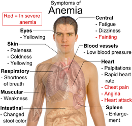
SYMPTOMS
Children with DBA appear to be normal, healthy infants at birth.
DBA have pregnancy-induced hypertension, premature separation of the placenta, or a breech presentation. Intrauterine growth retardation has been found in more than 10% of reported patients. Pregnancy and birth histories are abnormal in approximately 10% of these mothers.
Approximately 40% of the patients present with one or more congenital defects. Most of these abnormalities belong to the following categories:
- craniofacial dysmorphism including microcephaly, congenital cataract or glaucoma, strabismus, and a high-arched palate or cleft palate;
- prenatal or postnatal growth failure, independent of steroid therapy;
- neck anomalies (eg, fusion of the cervical vertebrae with flaring of the trapezius muscle - Klippel-Feil syndrome - giving a Turner syndrome appearance);
- thumb malformations;
- weak or absent radial pulses.
Certain facial characteristics often appear in children with DBA. The facial characteristics may consist of tow-colored hair (extremely blonde, almost white), a snub nose, wide-set eyes, and thick upper lip. In addition, the head can be small with almond-shaped eyes and a pointed chin. Various other anomalies can also be observed in children with DBA, such as horseshoe kidneys, duplication of ureters, ventricular and atrial septal defects, hypogonadism, ear malformations, blue sclera, mental retardation, congenital hip dislocation, or tracheoesophageal fistula.

DIAGNOSIS
The diagnosis is based on the presence of:
- normochromic, usually macrocytic, anemia;
- reticulocytopenia;
- normal or slightly decreased leukocyte counts;
- normal or increased platelet counts;
- normocellular bone marrow with selective deficiency of red cell precursors.
DBA has been associated with mutations in nine genes that encode ribosomal proteins. A mutation in one of these nine genes is identified in approximately 53% of individuals with DBA. Such testing is available clinically.
Clinical Diagnosis
The diagnosis of classic DBA, is made when all four of the following diagnostic criteria are met [Vlachos et al 2008, Vlachos & Muir 2010]:
- age younger than one year;
- macrocytic anemia with no other significant cytopenias;
- reticulocytopenia;
- normal marrow cellularity with a paucity of erythroid precursors.
The following laboratory findings are observed in most, but not all, individuals with DBA [Glader & Backer 1988, Willig et al 1999b]:
- Increased red cell mean corpuscular volume (MCV)
- Elevated erythrocyte adenosine deaminase activity (eADA) (observed in 80%-85%) [Glader & Backer 1988, Vlachos et al 2008]
- Elevated hemoglobin F (HbF) concentration
Other findings include:
- Growth retardation (observed in 30%)
- Congenital malformations (observed in ~30%-50%), in particular craniofacial, upper-limb, heart, and genitourinary malformations [Ball et al 1996, Willig et al 1999b, Lipton et al 2006]
The following are major supporting diagnostic criteria:
- Identification of a disease-causing mutation in one of the genes known to be associated with DBA
- Family history consistent with autosomal dominant inheritance
The following are minor supporting diagnostic criteria:
- Elevated eADA
- Elevated HbF concentration
- One or more congenital anomalies described in classic DBA
- No evidence of another inherited disorder of bone marrow function (see Differential Diagnosis) [Vlachos et al 2008]
If the family history is positive for other individuals with DBA and the family-specific mutation is known, any relative with the family-specific mutation should be considered to have non-classic DBA regardless of the presence of phenotypic findings.
Within families, some affected individuals may exhibit “non-classic” DBA, which is characterized by:
- Mild or absent anemia with only subtle indications of erythroid abnormalities such as macrocytosis, elevated eADA, and/or elevated HbF concentration
- Onset later in life [Willig et al 1999b, Lipton et al 2006]
- A phenotypically normal parent of an affected offspring
- Congenital anomalies or short stature consistent with DBA and minimal or no evidence of abnormal erythropoiesis [Lipton & Ellis 2010]
Persons with simplex cases (i.e., a single occurrence in a family) who do not meet clinical diagnostic criteria, but who have a disease-causing mutation in one of the genes known to be associated with DBA, should be considered to have non-classic DBA [Vlachos et al 2008].
Testing
Bone marrow aspirate shows normocellular bone marrow with:
- Erythroid hypoplasia;
- Marked reduction in normoblasts;
- Persistence of pronormoblasts on occasion;
- Normal myeloid precursors and megakaryocytes.
Molecular Genetic Testing
DBA has been associated with mutations in the nine following genes that encode ribosomal proteins. A mutation in one of these nine genes is identified in approximately 53% of individuals with DBA: RPS19, RPL5, RPL11, RPL35A, RPS24, RPS17, RPS7, RPS10, RPS26 [Doherty et al 2010].
PATHOGENESIS
Molecular Genetic Pathogenesis
Haploinsufficiency of RPS19 and RPS24 has been shown to be the basis for DBA. In a subset of affected individuals, frameshift mutations lead to degradation of the mutated transcripts [Hamaguchi et al 2002, Campagnoli et al 2004, Gazda et al 2004, Gazda et al 2006]. Recent studies have also demonstrated that DBA-causing missense RPS19 mutations affect RPS19 conformation and stability, triggering proteasome-mediated degradation or blocking its incorporation into pre-ribosomes [Angelini et al 2007, Gregory et al 2007, Kuramitsu et al 2008].

CAUSES AND RISK FACTORS
Some people have a family history of the disorder. More than half of people with DBA have a known genetic cause. In many people with DBA, doctors do not know the cause. If someone has DBA there is up to a 50% chance that each of his or her children will have DBA.
THERAPY
Eventually, 40% of individuals are steroid dependent, 40% are transfusion dependent, and 20% go into remission [Chen et al 2005, Vlachos et al 2008].
- Corticosteroids administration. Corticosteroids can initially improve the red blood count in approximately 80% of affected individuals.
The recommended corticosteroid is prednisone with a starting dose of 2 mg/kg/day given orally once a day in the morning, beginning when the child is at least six months old. An increase in hemoglobin concentration is usually seen in two to four weeks.
Corticosteroids may be slowly tapered to the minimal effective dose. Monitoring of blood counts is needed to ensure that the red cell hemoglobin concentration remains at 80-100 g/L, the minimum required for transfusion independence.
The corticosteroid maintenance dose varies and can be extremely low in some individuals. The recommended maximum maintenance dose is ≤0.5mg/kg/day or ≤1 mg/kg every other day.
If the recommended steroid dose cannot sustain the red cell hemoglobin concentration in an acceptable range (usually one month), the corticosteroids should be tapered and discontinued.
- Red blood cell transfusion. If the individual is resistant to corticosteroid therapy, chronic transfusion with packed red blood cells is necessary. The goal of transfusion therapy is a red cell hemoglobin concentration of 80-100 g/L, which is usually adequate for maintaining growth and development [Vlachos et al 2008, Vlachos & Muir 2010].
Hematopoietic stem cell transplantation (HSCT) is the only curative therapy for DBA. Persons with DBA who are transfusion-dependent or develop other cytopenias are often treated with HSCT.
- Bone Marrow Transplant. At present, this is the only cure for DBA. Bone marrow transplant involves the replacement of
diseased bone marrow with another person's healthy bone marrow. The decision to proceed with bone marrow transplant
should be discussed with your child's Hematologist and a Bone Marrow Transplant Team.
- Cancer treatment. Treatment of malignancies should be coordinated by an oncologist.
Prevention of Secondary Complications
Transfusion iron overload is the most common complication in transfusion-dependent individuals. The following methods are used both to assess for evidence of transfusion iron overload and to evaluate the effectiveness of iron chelation therapy:
- Measurement of iron concentration in a liver biopsy specimen, which accurately determines total body iron accumulation;
- MRI for assessing iron loading in the liver and heart;
- Magnetic biosusceptometry (SQUID), which gives a measurement of hepatic iron concentration
Note: Although the latter two methods of total iron measurement are noninvasive, they are available at only a limited number of centers and should be correlated with the “gold standard” liver biopsy [Cappellini & Piga 2008, Vlachos et al 2008];
- Deferasirox is recommended in individuals age two years or older. It is administered once daily in an oral dose of 20-30 mg/kg/day.
[Cappellini & Piga 2008, Porter et al 2008, Vlachos et al 2008];
- Desferrioxamine is administered four to seven nights a week in an eight- to 12-hour subcutaneous infusion via a portable pump. The recommended initial dose is 40 mg/kg/day; the maximum dose is 50-60 mg/kg/day. [Cappellini & Piga 2008, Vlachos et al 2008].
DIFFERENTIAL DIAGNOSIS
A diagnosis of DBA should be accepted conditionally if its onset occurs after the age of 2 years. In this case, the notion of similar cases in the family, elevated erythrocyte adenosine deaminase alone in the patient or apparently healthy family members, potentially associated malformations in the proband should be taken into account as well.
The main differential diagnosis is Parvovirus B19 erythroblastopenia in its neonatal form, acquired in utero during a primary maternal infection. This possibility must be excluded in all cases by a negative polymerase chain reaction (PCR) search for parvovirus B19 DNA in bone-marrow hematopoietic cells.
The diagnosis of Fanconi anemia can more rarely be considered. It is a congenital medullary aplasia discovered during the first years of life and associated with small stature and a variable setting of malformation(s). Should doubt persist, the search for spontaneous and provoked (by caryolysin or diepoxybutane) in vitro chromosome fragility of circulating lymphocytes will allow the distinction to be made. The chromosomal fragility typical of Fanconi’s anemia is not found in DBA.
Transitory erythroblastopenia of childhood is an acquired disease, probably of infectious origin, occurring often as limited outbreaks. It occurs later than DBA, most frequently after 2 years of age. It is not associated with malformations; there is no family history of the disease; it resolves spontaneously; no rise of erythrocyte adenosine deaminase is observed.
Autoimmune erythroblastopenia, rare in children, is acquired and most often occurs in a setting of dysimmunity (autoimmune hemolytic anemia, in particular).
Congenital dyserythropoieses are inherited aregenerative anemias. However, analysis of the myelogram detects no quantitative deficit of the erythroid cell line but rather qualitative anomalies of the maturation of this cell line.

CONCLUSION
Corticosteroids and chelation therapy have increased the life expectancy and an improved quality of life for children with DBA. The quality of life is improving for patients who respond to corticosteroid therapy. Chelating agents have also improved the quality of life for those who do not respond to corticosteroid therapy. As advancements occur in gene and cord blood stem cell research, the quality of life and life expectancy of patients with DBA will hopefully increase. Until those advancements are reality, nursing must help those patients who transition from pediatrics to young adult- hood make that transition as smooth as possible. Nurses may find that patients with DBA who develop cardiovascular diseases and malignancies are frequently patients in the intensive care unit; nurses must be sensitive to the psychosocial aspects of care, as well as the physiological aspects of care. Intimacy versus isolation can be a difficult stage to handle under normal circum- stances and even more difficult in the face of a chronic illness. This is a time when life-long relationships often develop. Support groups can be helpful and help broaden the patient’s circle of friends and contacts.
