INTRODUCTION
Myostatin, or growth differentiation factor 8, is a protein that in humans is encoded by the MSTN gene and is a secreted growth differentiation factor that is a member of the TGF beta protein family. The TGF beta protein family controls primarily proliferation and cellular differentiation; myostatin inhibits muscle differentiation and growth in the process known as myogenesis and is produced primarily in skeletal muscle cells, circulates in the blood and acts on muscle tissue.
The MSTN gene encoding myostatin was discovered in 1997 by geneticists Dr. Se-Jin Lee and Alexandra McPherron who also produced a strain of mutant mice that lack the gene, which had twice as much muscle as normal mice. Like other TGF-β family members, myostatin is synthesized as a precursor protein, the Myostatin Propeptide Human which is a 27.8 kDa protein containing 244 amino acid residues of the human Myostatin Propeptide; the propeptide undergoes proteolytic processing at a dibasic site to generate an N-terminal propeptide and a disulfide-linked C-terminal dimer, which is the biologically active molecule. The circulating form of myostatin consists of a latent complex of the myostatin C-terminal dimer and other proteins, including the myostatin propeptide, which inhibit the biological activity of the C-terminal dimer. The enzyme that cleavages the myostatin propeptide is unknown, but some researchers suggest that members of the bone morphogenetic protein-1/tolloid (BMP-1/TLD) family of metalloproteinases may be involved in activating latent myostatin in vivo. Human myostatin consists of two identical subunits, each consisting of 109 amino acid residues. Its total molecular weight is 25.0 kDa. Myostatin displays the traditional TGF-β family hand-shaped architecture, with each monomer consisting of four curved beta strands or ‘fingers', a cystine knot motif in the ‘palm' region, and a major helix or ‘wrist'.

Surface representation of the electrostatic potential of Fst288 in complex with myostatin. Surfaces are coloured by potential on the solvent accessible surface on a scale of −12.5 to 12.5 kbT/ec (red to blue)
Regulation of skeletal muscle mass in mice by a new TGF-p superfamily member, 1997
The structure of myostatin:follistatin 288: insights into receptor utilization and heparin binding, 2009
Activation of latent myostatin by the BMP-1/tolloid family of metalloproteinases, 2003
MYOSTATIN SIGNALING PATHWAY AND REGULATION
Myostatin binds to the activin type II receptor, resulting in a recruitment of a coreceptor called Alk-3 or Alk-4. The Activin type 2 receptors modulate signals for other ligands belonging to the Transforming growth factor beta superfamily of ligands, like Activin, Inhibin, Bone morphogenetic proteins and Nodal. They are all involved in a host of physiological processes including, growth, cell differentiation, homeostasis, osteogenesis, apoptosis and many other functions. Myostatin binds to ACVR2B and to a lesser extent ACVR2A. In mice that were ACVR2A / (null) mutants there was an increase in muscles’ size.
Although the role of myostatin in regulating muscle mass is well established, the molecular mechanisms mediating the response are not fully understood. Myostatin receptor activates the Smad2/3 signal transduction pathways which inhibit activation of the Akt/mTOR/p70S6 protein synthesis pathway, which mediates both differentiation in myoblasts and hypertrophy in myotubes. Myostatin receptor activates also P38 MAPK pathway that are responsive to stress stimuli and are involved in cell differentiation, apoptosis and autophagy.
However, rather than causing upregulation of the E3 ubiquitin ligases muscle RING-finger 1 (MuRF1) and muscle atrophy F-box (MAFbx), previously shown to mediate skeletal muscle atrophy, myostatin decreases expression of these atrophy markers in differentiated myotubes, as well as other genes normally upregulated during differentiation. These findings demonstrate that myostatin signaling acts by blocking genes induced during differentiation, even in a myotube, as opposed to activating the distinct “atrophy program”.

Myostatin signaling pathway
Myostatin promoter activity was also upregulated by TGF-β and members of the SMAD transcription factor family; this suggests that myostatin may autoregulate its own expression through activation of SMADs.
Also FoxO1, a transcription factor insulin-regulated, increased myostatin mRNA expression and promoter activity.
The proinflammatory cytokine TNF-α has the potency to induce the expression of myostatin in C2C12 skeletal muscle myocytes via the activation of p38MAPK and NF-κB.
In Silver Sea Bream GH caused significant decrease of MSTN1 transcript in white muscle but elevated the abundance of this transcript in red muscle. Injection of 11KT and cortisol resulted in decreased MSTN1 mRNA in red muscle whereas the abundance of MSTN2 mRNA remained relatively unchanged following hormone administration.
Although these findings the signaling ways that modifies myostatin gene expression is still unclear.
Myostatin reduces Akt/TORC1/p70S6K signaling, inhibiting myoblast differentiation and myotube size, 2009
Regulation of myostatin expression and myoblast differentiation by FoxO and SMAD transcription factors, 2007
Impact of exercise training on myostatin expression in the myocardium and skeletal muscle in a chronic heart failure model, 2009
Hormonal Regulation of Myostatin Expression in Silver Sea Bream Muscle, 2011
MYOSTATIN DEFICIENCY
Animals’ model lacking of myostatin or treated with substances that block the activity of myostatin have significantly larger muscles, as proved on mice.
It’s still not clear if the muscles grow is lead by hypertrophy, hyperplasia or both because different tests on different animal model produced conflicting results: the Double-muscled Piedmontese cattle have a C313Y mutation in myostatin and show increased skeletal muscle mass which resulted from an increase of myofiber number (hyperplasia) without that of myofiber size (hypertrophy), but transgenic mice exhibited dramatic increases in the skeletal muscle mass resulting from hyperplasia without hypertrophy. These results suggest that the myostatin containing the missense mutation exhibits a dominant negative activity and that there are two types in the dominant negative form of myostatin, causing either hypertrophy or hyperplasia.
Some studies indicate that myostatin inhibition in healthy mice causes an increase muscle fiber size without increasing force production, although myostatin increases atrogin-1 gene (and increased protein ubiquitination) in mice’s myotubes, but not in human myotubes.
However, treatment of mdx mice, a murine model of Duchenne muscular dystrophy, with the propeptide of myostatin, increased both Fo and sFo of EDL muscles.
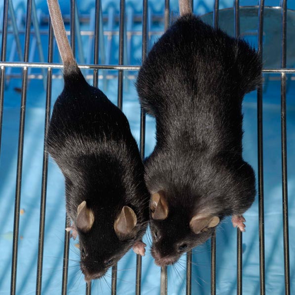
Comparison between a wild type mouse and mice mstn / (a mice with homozygous mutation in myostatin)
In medical literature there are really few cases of myostatin pathway’s mutations in human. There is a study of the New England Journal of Medicine about a newborn with has a loss-of-function mutation in the myostatin gene diagnosed by encoding his DNA. Despite the mutations the children have no medical issue.
So mutations that cause a reduction of the pathway’s activation can not be all detected because it probably lead only to a bigger muscle size without evident other effects. However, a technique for detecting mutations in myostatin variants has been developed.

Photographs of the Child at the Ages of Six Days and Seven Months
Decreased specific force and power production of muscle fibers from myostatin-deficient mice are associated with a suppression of protein degradation, 2011
A missense mutant myostatin causes hyperplasia without hypertrophy in the mouse muscle, 2004
Myostatin Mutation Associated with Gross Muscle Hypertrophy in a Child, 2004
MYOSTATIN INHIBITOR THERAPY
Blocking the activity of myostatin may have therapeutic application in treating muscle wasting diseases considering the great muscle gains in animal model.
1. CACHEXIA
Cachexia, progressive loss of fat and muscle mass despite adequate nutrition, is a devastating complication of cancer associated with poor quality of life and increased mortality. In an experimental model of cancer cachexia is associated with modulations of myostatin signalling and maybe the cytokine tumour necrosis factor-alpha may be relevant in this regard. A recent research studies how myostatin inhibition might influence cancer cachexia using genetic and pharmacological approaches. First, hypermuscular myostatin null mice were injected with Lewis lung carcinoma or B16F10 melanoma cells. Myostatin null mice were more sensitive to tumor-induced cachexia, losing more absolute mass and proportionately more muscle mass than wild-type mice because myostatin null mice lack expression from development.
However, the Trichostatin A (histone deacetylase inhibitor) has been shown to increase muscle mass in normal and dystrophic mice by inducing the myostatin inhibitor, follistatin. Although Trichostatin A administration induced muscle growth in normal mice, it failed to preserve muscle in colon-26 cancer cachexia.
Systemic administration of ACVR2B-Fc, Activin receptor extracellular domain/Fc fusion protein, potently inhibited muscle wasting and protected adipose stores in both colon-26 and Lewis lung carcinoma cachexia, without affecting tumor growth. Enhanced cachexia in myostatin knockouts indicates that host-derived myostatin is not the sole mediator of muscle wasting in cancer. More importantly, skeletal muscle preservation with ACVR2B-Fc establishes that targeting myostatin-family ligands using ACVR2B-Fc or related molecules is an important and potent therapeutic avenue in cancer cachexia.

ACVR2B-Fc pathway
Acute inhibition of myostatin-family proteins preserves skeletal muscle in mouse models of cancer cachexia, 2010
2. SARCOPENIA
The progressive loss of skeletal muscle mass, strength and/or function with advancing age, termed sarcopenia, poses a major threat to independence and quality of life. Therefore, there is significant merit in better understanding the biology of sarcopenia and developing therapeutic interventions to prevent, slow or reverse its progression. Since the discovery of myostatin, there has been great interest in it as a potential mediator of sarcopenia as well as a therapeutic target. In a recent study a fusion protein consisting of the extracellular ligand-binding domain of activin type IIB receptor with the Fc portion of human immunoglobulin G (ActRIIB-Fc) was used to inhibit signaling through this pathway in adult, 18-month-old, and orchidectomized mice. Significant muscle growth and enhanced muscle function were observed in adult mice treated for 3 days with ActRIIB-Fc. The ActRIIB-Fc-treated mice had enhanced fast fatigable muscle function, with only minor enhancement of fatigue-resistant fiber function. The ActRIIB-Fc-treated 18-month-old mice and orchidectomized mice showed significantly improved muscle function. Treatment with ActRIIB-Fc also increased bone mineral density and serum levels of a marker of bone formation. These observations highlight the potential of targeting ActRIIB receptor to treat age-related and hypogonadism-associated musculoskeletal degeneration.
Increased muscle force production and bone mineral density in ActRIIB-Fc-treated mature rodents, 2013
Myostatin and Sarcopenia: Opportunities and Challenges - A Mini-Review, 2014
3. DYSTROPHY
Myostatin has also shown promising results as a potential therapeutic target for treating muscular dystrophy in animal studies. Muscular dystrophy is an inherited disorder involving progressive muscle weakness and wasting. Duchenne’s muscular dystrophy (DMD) is the most common X-linked neuromuscular disease and is estimated to affect 1 in 3500 newborn males. DMD is characterized by progressive and severe muscle loss that leads to loss of ambulation, with those affected often becoming wheelchair dependent toward the end of the first decade of life. The disease is caused by mutations in the DMD gene resulting in quantitative and/or qualitative disturbances in expression of the gene product, dystrophin. Dystrophin is associated with the membrane-bound dystroglycan complex (DGC), which forms an important link with laminin, a constituent of the extracellular matrix. Mutations in the genes encoding various members of the complex are thought to disrupt sarcolemmal integrity, resulting in a variety of X-linked and limb girdle muscular dystrophies. Although DMD remains incurable, steady advances using gene-based, cell-based, and pharmacological strategies in experimental models of the disease continue to be made.
Myostatin blockade therefore offers a strategy for reversing muscle wasting in Duchenne’s muscular dystrophy (DMD) without resorting to genetic manipulation. The pharmacological blockade using a myostatin propeptide stabilized by fusion to IgG-Fc improved pathophysiology of the mdx mouse model of DMD.

Increase of muscle size by propeptide-mediated blockade in mdx mice. Treated mdx mice (red bars) had larger CSA (a); 1.8 ± 0.5 vs. 1.4 ± 0.8 mm2
Myostatin propeptide-mediated amelioration of dystrophic pathophysiology, 2004
HUMAN MYOSTATIN INHIBITOR DRUGS
As of 2014, no myostatin-inhibiting drugs for humans are on the market, but an antibody genetically engineered to neutralize myostatin was developed by New Jersey pharmaceutical company Wyeth. Stamulumab (MYO-029) is an experimental myostatin inhibiting drug developed for the treatment of muscular dystrophy (MD). Stamulumab was formulated and tested by Wyeth in Collegeville, Pennsylvania. Stamulumab is a G1 immunoglobulin antibody which binds to myostatin and prevents it from binding to its target site, thus inhibiting the growth-limiting action of myostatin on muscle tissue. Research completed in 2002 found that Stamulumab might one day prove to be an effective treatment for Duchenne muscular dystroph.
Wyeth undertook a Phase 1 and 2 clinical trial in 2005 and 2006 of stamulumab. The multiple ascending dose trial (36 patients per cohort) contained some measures of efficacy. The trial's participants included people afflicted with Facioscapulohumeral muscular dystrophy, Becker's muscular dystrophy, and Limb-girdle muscular dystrophy. In January 24, 2008, Wyeth announced that the study had been accepted by a peer-reviewed journal and publication was expected "in the next few months". The publication appeared in Annals of Neurology in May 2008. This trial supports the hypothesis that systemic administration of myostatin inhibitors provides an adequate safety margin for clinical studies, but here were no improvements noted in exploratory end points of muscle strength or function (but the study was not powered to look for efficacy). However, on 11 March 2008 it was announced that Wyeth would not develop the drug further for MD, but would continue to explore myostatin inhibition along with other strategies.
A phase I/IItrial of MYO-029 in adult subjects with muscular dystrophy, 2008
CONCLUSION
There has been progress in evaluating antimyostatin therapies in animal models of muscle wasting disorders; some programs have progressed into clinical development. In normal mice myostatin deficiency results in enlarged muscles with increased total force but decreased specific force (total force/total mass). An increase in myofibrillar protein synthesis without concomitant satellite cell proliferation and fusion leads to muscle hypertrophy with unchanged myonuclear number. A specific force reduction is not observed when atrophied muscle, the predominant therapeutic target of myostatin inhibitor therapy, is made myostatindeficient.
Myostatin inhibition remains a promising therapeutic strategy for a range of muscle wasting disorders.
Myostatin inhibitors as therapies for muscle wasting associated with cancer and other disorders, 2013
Filippo Palmesino