Francesco Riscaldino
Luca Roggero
INTRODUCTION
Paracetamol: therapeutic indications
Paracetamol, or acetaminophen, or APAP, chemically named N-acetyl-p-aminophenol, is a widely used over-the-counter analgesic and antipyretic.
Paracetamol is classified as a mild analgesic. It is commonly used for the relief of headaches and other minor aches and pains and is a major ingredient in numerous cold and flu remedies. In combination with opioid analgesics, paracetamol can also be used in the management of more severe pain such as post-surgical pain. Though paracetamol is used to treat inflammatory pain, it is not generally classified as an NSAID (non-steroidal anti-inflammatory drug) because it exhibits only weak anti-inflammatory activity.
Paracetamol: overdose and toxicity
While generally safe for use at recommended doses (1,000 mg per single dose and up to 4,000 mg per day for adults), even small overdoses can be fatal. Compared to other over-the-counter pain relievers, paracetamol is significantly more toxic in overdose but may be less toxic when used chronically at recommended doses.
Dunaliella salina in Paracetamol overdose
Dunaliella salina, an unicellular green algae which is one of the richest natural producers of carotenoid, was investigated for hepatoprotective and antioxidant activity against paracetamol-induced liver damage in rats and exhibited a potent hepatoprotective effect, which may be due to both the increase of antioxidant enzymes activity and inhibition of lipid peroxidation.
Acetaminophen, Drug.com
"Hepatoprotective and antioxidant activity of Dunaliella salina in Paracetamol-induced Acute Toxicity in Rats, Indian Journal of Pharmaceutical Sciences; Nov 2013"
CHARACTERISTICS OF PARACETAMOL
Mechanism of action
Paracetamol: pharmacokinetics
Metabolism
Paracetamol: molecular mechanism
Toxicity
Paracetamol: side effects and toxicity
DUNALIELLA SALINA

Taxonomy, life-history and morphology
Dunaliella is a unicellular, bi-flagellate, naked green alga. The genus was first described by Teodoresco with the type of species being Dunaliella salina, and at present a total of 29 species, as well as a number of varieties and forms, are recognized.
Dunaliella is morphologically similar to Chlamydomonas, with the main difference being the absence of a cell wall in Dunaliella. Dunaliella has two flagella of equal length and a single, cup-shaped chloroplast.
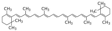
In D. salina the chloroplast accumulates large quantities of B-carotene so that the cells appear orange-red rather than green. The carotenoid are in the form of droplets (plastoglobuli) located at the chloroplast periphery and consist of a mixture of the cis- and transisomers of ß-carotene. A typical composition, expressed as percentages of total ß-carotene is 15-cis-ß-carotene, 10%; 9-cis-ß-carotene, 41%; all-trans-ß-carotene, 42%; other isomers, 6%.
In the alga this ß-carotene seems to act as photo-protective ‘sun-screen’ to protect the chlorophyll and the cell DNA from the high irradiance which characterizes the normal habitat of D. salina. The ß-carotene also acts as a ‘carbon sink’ to store the excess carbon produced during photosynthesis under conditions where growth is limited but photosynthetic carbon fixation must continue.

Cell shape in this genus is very variable, being oval, spherical, cylindrical, ellipsoidal, egg-, pear- or spindle-shaped with radial, bilateral or dorsoventral symmetry or being asymmetrical. Cells in any given species may change shape with changing conditions, often becoming spherical under unfavorable conditions. Cell size also varies with growth conditions and light intensity.
The cells divide by lengthwise division in the motile state. Under certain conditions they may also develop into a palmella stage and become embedded in a thick layer of mucilage, or they may form aplanospores with a thick, rough wall. Sexual reproduction is rarely observed in cultures, but more often in the field, and it is by isogamy, with conjugation proceeding.The zygote is green or red and is surrounded by a very thick, smooth wall of sporopollenin.
After a resting stage the zygote nucleus divides meiotically, forming up to 32 cells which are liberated through a rupture in the mother cell wall.
"The mass culture of Dunaliella salina", 1990
Ecology
The genus Dunaliella has marine and halophilic representatives. Freshwater species have also been described although it has been proposed that these should be assigned to a different genus. Dunaliella also has a very wide pH tolerance ranging from pH 1 (D. acidophila) to pH 11 (D. salina). In fact, D. salina is one of the most environmentally tolerant eukaryotic organisms known and can cope with a salinity range from seawater (= 3% NaCl) to NaCl saturation
(= 31% NaCl), and a temperature range from <0 °C to >38 °C. The optimum growth temperature for D. salina is in the range of 20 to 40°C depending on the strain. Dunaliella salina can tolerate extremely low temperatures to below freezing, but temperatures greater than 40°C are usually lethal. There is also a strong interaction between the growth rate, temperature and salinity and between light intensity and temperature tolerance.
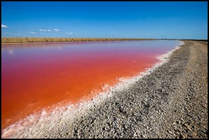
Dunaliella species are commonly observed in salt lakes in all parts of the world from tropical to temperate to polar regions where they often impart an orange-red colour to the water. Marine Dunaliella species can generally be isolated from seawater, although they are not very abundant in nature.
"The mass culture of Dunaliella salina", 1990
The physiology of carotenogenesis
Dunaliella salina is characterized by its ability to accumulate very high concentrations of β-carotene. Concentrations of up to 14% of dry weight have been reported. The halophilic species D. parva also accumulates high concentrations of β-carotene (<4% of dry weight), whereas none of the other species accumulate such large amounts. Recently, what appears to be a new species of Dunaliella has been isolated in Chile which accumulates not only high concentrations of β-carotene, but also a-carotene. High levels of β-carotene accumulation require high salinity, high temperature and high light. Nutrient limitation, especially N limitation, also enhances carotenoid formation.
In general, carotenogenesis is greatest under sub-optimal growth conditions when the specific growth rate is low.
Light is essential for carotenogenesis, but the maximum level of β-carotene in the cell is dependent on the salinity. Carotenoid formation is rapid. For example, when the salinity of a culture of D. salina is increased from 15 to 25% NaCl the total carotenoid increases linearly from < 10 to 260 mg (g cell protein)-1 over a period of 4 to 5 days. The proportions of β-carotene increases from 50% to 90% during this period whereas there was no measurable increase in the other carotenoid, α-carotene, lutein and zeaxanthin. Fortunately, from the mass culture point of view, the cell content of Bcarotene does not decrease at the same rapid rate when the salinity of the medium is reduced as occurs in outdoor cultures when it rains. When carotenoid-rich cells are transferred from a high salinity (25% NaCl) medium to a low salinity (15% NaCl) medium, there is only a gradual decline in cell carotenoid content over a period of several weeks. Most of this decline is not due to β-carotene breakdown, but rather due to reapportionment of the β-carotene as the cells divide.
"The mass culture of Dunaliella salina", 1990
β-carotene applications
Until today, β-carotene remains the major natural product harvested from D. salina.
Common uses of β-carotene include food colouring, additives to multivitamin preparations, health-food products, cosmetics and as a precursor of vitamin A. The potential ability of carotenoids to act as antioxidants and immunomodulatory agents has led to more active research investigating their application for the prevention of human cancers. Therefore, D. salina could be used as a source of antioxidant to improve free radical scavenging activities in the body and protect cells from oxidative damage.
"Hepatoprotective and antioxidant activity of Dunaliella salina in Paracetamol-induced Acute Toxicity in Rats, Indian Journal of Pharmaceutical Sciences; Nov 2013"
"The mass culture of Dunaliella salina", 1990
HEPATOPROTECTIVE AND ANTIOXIDANT ACTIVITY OF DUNALIELLA SALINA
In recent years, attention has been focused upon the role of biotransformation of chemicals to highly reactive metabolites that initiate cellular toxicity. Several chemicals, including clinically used drugs, can cause cellular damage through metabolic activation of the chemical to highly reactive compounds such as free radicals, carbenes and nitrenes. Paracetamol, the widely used analgesic antipyretic drug, though considered a safe drug, it produces hepatic necrosis and renal failure when given in high doses. Oxidative stress was reported to play a fundamental role in the pathogenesis of paracetamol-induced liver damage.
In acute toxicity study, LD50 was estimated to be >5000 mg/kg. Hence, one-fifth and one-tenth of the LD50, (500 and 1000 mg/kg) were selected to evaluate the effect of D. salina against praracetamol-induced liver injury in the study.
ALT and AST as index of liver cells damage
The liver enzymes, AST and ALT are cytoplasmic in origin and released into the blood after hepatic cell damage. However, ALT is more specific to the liver and is deemed a better parameter for detecting liver injury because high level of AST indicates liver damage as well as cardiac infarction and muscle injury. Furthermore, serum ALP and bilirubin levels are related to the function of hepatic cell. Increase in ALP level is due to increased synthesis, in the presence of increasing biliary pressure.
Pretreatment with Dunaliella salina
In agreement with previous studies, the animals treated with a dose of 3 g/kg of paracetamol showed a significant hepatic damage at 24 h after dosing, as elicited by the significant (P<0.05) elevated levels of hepato-specific serum markers, AST, ALT and ALP as well as total and direct bilirubin. Pretreatment with D. salina extract was protective, as indicated by significant (P<0.05) reduction of all the previous parameters. The normalisation of the above enzyme levels in rats treated with the algal dose of 1000 mg/kg was comparable with that observed for silymarin, which clearly establishes the hepatoprotective effect of this dose. This indicates that administration of D. salina extract at the high dose might be able to induce regeneration of liver cells, reducing the leakage of the above enzymes into the blood.

SOD forms a crucial part of the cellular antioxidant defense mechanism. It removes superoxide (O2−) by converting it to H2O2, which can be rapidly converted to water by catalase and glutathione peroxide (GPx). It is also known that MDA is one of the end products in the lipid peroxidation process. However, oxidative stress results in toxicity when the generated free radicals exceed the cell's capacity for their removal. In the present study, the increase in MDA and decrease in both TAC and SOD levels in rats administrated with paracetamol suggest enhancement of lipid peroxidation leading to tissue damage and failure of antioxidant defense mechanisms to prevent the formation of excessive free radicals. Treatment with D. salina extract significantly reversed these changes.
Alteration of biomembrane lipid profile disturbs its fluidity and increases microviscosity of the membrane as a result of cholesterol increasing, which leads to cellular rigidity. NO is a highly reactive oxidant produced by liver parenchymal and nonparenchymal cells from L-arginine via an inducible form of NO synthase. Overproduction of NO in the liver has been implicated as an important event in endotoxin shock and in other models of hepatic inflammation and injury. Intoxication of rats with paracetamol may have altered membrane structure and function as suggested by the increases in cholesterol and NO. However, pretreatment of rats with extracts of D. salina inhibited the alteration of lipid membranes and hence prevented alterations in the levels of cholesterol and NO. These results suggest that methanol extract of D. salina play a role in peroxidation by inhibiting free radical attacks on biomembranes.
Histological sections of liver showed that centrilobular necrosis, the pathognomonic feature of hepatotoxicity, which appeared in paracetamol-intoxicated rats, was strikingly reduced in D. salina pretreated rats. Furthermore, the congestion and inflammatory cell infiltration evoked by paracetamol was considerably decreased by D. salina indicating its possible antihepatotoxic action.

Effect of D. salina on the liver histopathological photomicrographs of the experimental groups of rats.
Histopathological photomicrographs (×400) of livers of various groups stained with haematoxylin and eosin. (a) Normal architecture of rat liver, (b) Necrosis and hepatocellular fatty degeneration (eccentric nuclei) in acetaminophen intoxicated liver and congestion of portal vein and peri-portal infiltration of inflammatory cells, © Lesser damage of hepatocytes and low index of necrosis (centrally located nuclei) in D. salina-500 mg/kg pretreated group, (d) Minimal damage of hepatocytes and very low index of necrosis in D. salina-1000 mg/kg pretreated group and mild congestion, (e) Very lesser damage of hepatocytes and low index of necrosis in silymarin pretreated group, narrow arrows refer to inflammatory cells infiltration, wide arrows refer to congestion of portal vein.
"Hepatoprotective and antioxidant activity of Dunaliella salina in Paracetamol-induced Acute Toxicity in Rats, Indian Journal of Pharmaceutical Sciences; Nov 2013"
CONCLUSION
In conclusion, D. salina extract could be considered as a potential source of natural antioxidant with hepatoprotective activity. Liver histopathology also showed that D. salina reduces the centrilobular necrosis, congestion and inflammatory cell infiltration evoked by paracetamol overdose. These results suggest that D. salina exhibits a potent hepatoprotective effect on paracetamol-induced liver damage in rats, which may be due to both the increase of antioxidant enzymes activity and inhibition of lipid peroxidation.
Further detailed investigations on this algae are needed in order to identify and isolate the hepatoprotective components in the extract and to justify its use in the treatment of liver disorder.
"Hepatoprotective and antioxidant activity of Dunaliella salina in Paracetamol-induced Acute Toxicity in Rats, Indian Journal of Pharmaceutical Sciences; Nov 2013"