Linkage betwen obesity and human genome

Obesity is a major public health issue.
Studies published in 2007 showed in humans that a chromosome region containing a gene called the Fat Mass and Obesity gene, FTO for short, was associated with increased body mass index.
The FTO gene was a candidate in the original studies because the DNA markers that showed the association with fat mass were within the FTO gene itself. However, these markers were within sequence that is not coding and that may be regulatory.
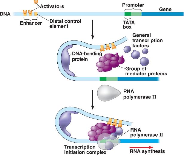
These sorts of regulatory regions can have effects over long distances and therefore on other genes.
The FTO gene has a role in determining BMI. It is also significant that increasing the expression of FTO increases food intake and is consistent with a number of human studies that measured food intake against people's DNA marker genotype in this region.
FTO is localized on chromosome 16.
Noncoding variation in single nucleotide polymorphisms (SNPs) within a 47-kilobase (kb) region of high linkage disequilibrium in introns 1 and 2 of FTO remains the strongest genetic association with risk to polygenic obesity and type 2 diabetes in humans.
Individuals homozygous for risk alleles of the associated SNPs weigh approximately 3 kg more than individuals homozygous for non-risk alleles, underscoring the significant phenotypic impact of these common variants.
IRX3 leads the way
Recently it has been discovered an important association betwen the risk of obesity and the expression a gene localizated on the same FTO chromosome, to about 1 megabase away, named IRX3.

The obesity-associated noncoding sequences within FTO are functionally connected with the homeobox geneIRX3.
There is no direct evidence that enhancers within the obesity-associated FTO introns are connected with regulation of FTO expression, infact quantitative trait locus (eQTL) analyses have systematically failed to show association between the obesity-associated SNPs and FTO expression in human tissues.
Protein Aminoacids Percentage
The Protein Aminoacids Percentage gives useful information on the local environment and the metabolic status of the cell (starvation, lack of essential AA, hypoxia)
Protein Aminoacids Percentage (Width 700 px) IRX3 and FTO

FTO cis-regulation
To chart directly the cis-regulatory circuitry within the FTO locus, it was used circular chromosome conformation capture followed by highthroughput sequencing (4C-seq).
4C-seq was carried out inwholemouse embryos (embryonic day 9.5; E9.5) and in adult (8 weeks) mouse brains, as previous work suggests that brain FTO expression modulates
metabolic parameters.
It was profiled the genomic interactions with promoters of genes located within a 1-megabase (Mb) window around the obesity-associated SNPs , including Fto and Rpgrip1l, and Irx3, a halfmegabase downstream.
There was a clear difference in interaction patterns among genes.
The Fto promoter chiefly participates in genomic interactions proximal to the gene promoter.
The interactions between Irx3 and the Fto obesity-associated region in adult mouse brains was confirmed with a test.
The promoter of Irx3 participates in numerous long-range interactions across a broad genomic region encompassing nearly 2 Mb, including *robust interactions with the obesity-associated interval within FTO, these data suggest that the obesity-associated interval is likely to be part of the regulatory
landscape of Irx3.*
It was next inferred that the long-range interactions between the obesity-associated FTO intron and IRX3 represent a conserved feature in vertebrate genomes.

The different genetic expression regulation between IRX3 e FTO
There are suggestion that the regulatory landscape of IRX3 spreads over megabase distances, whereas FTO expression is primarily regulated by regions proximal to its promoter.
To test this, it was engineered a human bacterial artificial chromosome (BAC) spanning 162 kb of the FTO locus, including its promoter and the 47-kb obesity-associated region.
It was recombineered a reporter cassette at the FTO translation start, and generated transgenic mice harbouring the engineered BAC. Transgenic mice expressed the reporter gene in multiple tissues, recapitulating the endogenous expression pattern of FTO.
It was next determined that a 1.2-kb region corresponding to the FTO promoter is sufficient to recapitulate most of the FTO expression pattern. In contrast, a 2.8-kb region corresponding
to the IRX3 promoter does not recapitulate any of the endogenous expression patterns of IRX3, suggesting that the broad expression patterns of FTO are primarily regulated by elements proximal to the promoter, and that IRX3 is endowed with an ancient, extensive cis-regulatory circuitry extending into FTO.
Evidence in humans
The gene expression studies in human brains corroborate the chromatin looping data, showing that the obesity-associated SNPs are associated with expression levels of IRX3, but not FTO.
In human brain samples FTO and IRX3 are highly expressed in multiple regions of the brain including cerebellum and hypothalamus.
Using a data set of 153 brain samples from individuals of European ancestry, represented by cerebellum, it was found significant association between 11 SNPs previously associated with increased body mass index (BMI), and expression of IRX3.
Data set of SNPs associated with BMI in 249,796 individuals, reveals that among SNPs significantly associated with IRX3 expression in brain or adipose tissue, only those associated with IRX3 in brain show highly significant associations with BMI.
IRX3 and his effects
There were used animal models to determine a potential role for IRX3 expression in the regulation of BMI and/or metabolism.
Micehomozygous for an Irx3-null allele (Irx3-knockout mice) are viable and fertile, with no evidence of embryonic lethality.It was observed a 25–30% reduction of body weight in Irx3-knockout mice compared to control littermates (wild type), independent of gender.
This difference becomes more pronounced if animals were subjected to a high-fat diet (HFD), with Irx3-knockout animals showing no significant body weight gain , contrasting to a 63% increase in control animals.

Furhermore, the percentage of fatmass in Irx3-knockout mice was significantly reduced without marked change of the lean mass ratio.
Irx3-knockout mice exhibit marked reduction in adiposity
with smaller fat depots as well as reduced adipocyte size. These results were confirmed by
differential gene expression of adiposity markers that is leptin, adiponectin and mcp1 in the perigonadal white adipose tissue (PWAT) of Irx3-knockout mice.
Importantly, Fto expression was not altered in the hypothalamus or PWAT of Irx3-knockout mice, suggesting that the lean phenotype of Irx3-knockout mice is not associated with Fto.

As Irx3-knockout mice were resistant to HFD-induced obesity and metabolic disorder, such as hepatosteatosis, we examined glucose homeostasis by performing glucose tolerance tests and insulin tolerance test at different time points in the presence or absence of HFD. In 8-week-old mice, no difference in GTT was found.
Although ageing and HFD led to glucose intolerance and insulin resistance in wild-type mice, Irx3-knockout mice showed none of these metabolic phenotypes.
Indirect calorimetric analysis showed higher energy expenditure in Irx3-knockoutmice.
Notable is that Irx3-knockout mice show upregulation of brown adipocyte markers in PWAT, including Ucp1, Ppargc1a, Prdm16 and cidea, as well as increased expression of Adrb3 encoding the b3-adrenergic receptor, suggestive of elevated sympathetic activation. It was also found a significant increase of Ucp1 expression in the brown adipose tissue (BAT) of Irx3-knockout mice.
Brown adipose tissue dissipates energy (lipids and glucose) and has been the focus for potential development of novel therapeutic strategies to treat obesity and diabetes. ‘Browning’ of White Adipose Tissue (WAT) by higher sympathetic tone and activation of BAT might lead to increased energy expenditure in Irx3-knockout mice and partially account for their leanness and protection from diet-induced obesity.
Browning of WAT by increased sympathetic activity is a phenomenon controlled by hypothalamic circuits integrating central and peripheral metabolism regulation.

Other data suggest that the obesity-associated SNPs are associated with IRX3 expression in brain.
It was determined that Irx3 is expressed in the arcuate nucleus and median eminence of the hypothalamus, two critical regions involved in the regulation of energy homeostasis.
| There are data that represent the first demonstration of the intersection of IRX3 biology with bodymass composition and metabolism, in fact previous work identified IRX3 overexpression in adipocytes as a hallmark of the molecular switch seen in patients after profound weight loss following bariatric surgery. |
(This evidence led us to ask how the gene is influential in the occurrence of obesity)
Future investigations will determine the precise molecular mechanisms by which IRX3 regulates metabolic parameters and this will lead to determinate possible new paths against obesity.
Obesity-associated variants within FTO form long-range functional connections with IRX3, 2014
Stefano Quaranta
Simone Silva