Signaling Pathways

Author: Gianpiero Pescarmona
Date: 19/09/2007

Description

The biological effects produced by intracellular actions are referred as intracrine

The biological effects produced by binding to cell surface receptors are called

  • autocrine - the cell signals itself through a chemical that it synthesizes and then responds to. Autocrine signaling can occur
    • by a secreted chemical interacting with receptors on the surface of the same cell
  • paracrine - chemical signals that diffuse into the area and interact with receptors on nearby cells. Examples are:
    • The release of cytokines that cause an inflammatory response in the area.
    • The release of neurotransmitters at synapses in the nervous system.
  • endocrine - the chemicals are secreted into the blood and carried by blood and tissue fluids to the cells they act upon.

The biological effects produced by interactions between cell membrane proteins are referred as iuxtacrine

Second Messengers

Second Messengers are molecules that relay signals received at receptors on the cell surface — such as the arrival of protein hormones, growth factors, etc. — to target molecules in the cytosol and/or nucleus.
They serve to greatly amplify the strength of the signal. Binding of a ligand to a single receptor at the cell surface may end up causing massive changes in the biochemical activities within the cell.

There are 3 major classes of second messengers:

1. cyclic nucleotides (e.g., cAMP and cGMP)
2. inositol trisphosphate (IP3) and diacylglycerol (DAG)
3. calcium ions (Ca2+)

Signalling molecules

effects depending on the origin of the signalling molecule

Modulation of the single steps of the signalling cascade and its bifurcations
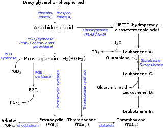
Arachidonic Acid

MAPK

Additional pathways

Cytohesins and centaurins control subcellular trafficking of macromolecular signaling complexes: Regulation by phosphoinositides and ADP-Ribosylation Factors 2002

Comments
2011-09-28T13:55:37 - Gianpiero Pescarmona

Cancer Res. 2011 Aug 18. [Epub ahead of print]
A mutation threshold distinguishes the anti- tumorigenic effects of the mitochondrial gene MTND1, an oncojanus function. 2011
Gasparre G, Kurelac I, Capristo M, Iommarini L, Ghelli A, Ceccarelli C, Nicoletti G, Nanni P, De Giovanni C, Scotlandi K, Betts CM, Carelli V, Lollini PL, Romeo G, Rugolo M, Porcelli AM.
Source
Dip. di Sc. Ginecologiche, Ostetriche e Pediatriche, Genetica Medica, Univ. di Bologna.
Abstract
The oncogenic versus suppressor roles of mitochondrial genes has been long debated. Peculiar features of mitochondrial genetics such as hetero/homoplasmy and mutation threshold are seldom taken into account in this debate. Mitochondrial DNA (mtDNA) mutations generally have been claimed to be pro-tumorigenic, but they are also hallmarks of mostly benign oncocytic tumors where they help reduce adaptation to hypoxia by destablizing hypoxia-inducible factor-1α (HIF1α). To determine the influence of a disassembling mtDNA mutation and its hetero/homoplasmy on tumorigenic and metastatic potential, we injected mice with tumor cells harboring different loads of the gene MTND1 m.3571insC. Cell cultures obtained from tumor xenografts were then analyzed to correlate energetic competence, apoptosis, α-ketoglutarate/succinate ratio and HIF1α stabilization with the mutation load. A threshold level for the anti-tumorigenic effect of MTND1 m.3571insC mutation was defined, above which tumor growth and invasiveness were reduced significantly. Notably, HIF1α destabilization and downregulation of HIF1α-dependent genes occurred in cells and tumors lacking Complex I, where there was an associated imbalance of α- ketoglutarate/succinate despite the presence of an actual hypoxic environment. These results strongly implicate mtDNA mutations as a cause of oncocytic transformation. Thus, the anti-tumorigenic and anti-metastatic effects of high loads of MTND1 m.3571insC, following complex I disassembly, defines a novel threshold- regulated class of cancer genes. We suggest these genes be termed oncojanus genes, to recognize their ability to contribute either oncogenic or suppressive functions in mitochrondrial settings during tumorigenesis.

Attachments
fileuserdate
VEGF.doc29230905/03/2008
AddThis Social Bookmark Button