Because the lipid bilayer of the eucaryotic plasma membrane is impermeable for hydrophilic molecules, glucose is transported across the plasma membrane by membrane associated carrier proteins, glucose transporters. There are 2 different types of transporter proteins, which mediate the transfer of glucose and other sugars through the lipid bilayer:
- Sodium-dependent glucose cotransporters (SGLT)
- Facilitative glucose transporters (GLUT).
The SGLT family
The SGLT family comprises Na+-dependent glucose co-transporters (SGLT1 and SGLT2), the glucose sensor SGLT3, the widely distributed inositol and multivitamin transporters SGLT4 and SGLT6,9 and the thyroid iodide transporter SGLT5.
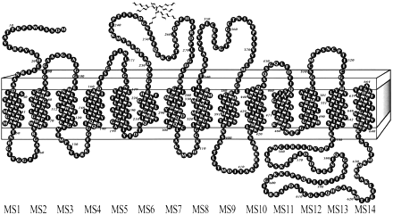
The sodium-dependent glucose transporters, also known as co-transporters or symporters, are integral membrane proteins (wich contains 14 membrane spanning α-helices with both the hydrophobic N-terminus and C-terminus facing the extracellular site)that mediate the transport of glucose and, with lower affinity, galactose across the plasma membrane by an active transport mechanism. This transport process is a cotransport of glucose molecules and sodium ions.
The transporters are expressed in different tissues:
- SGLT1 mainly in intestine, heart and kidney (proximal tubule of the nephron)
- SGLT2 mainly in kidney (proximal tubule of the nephron)
- SGLT3 in intestine, spleen, liver, kidney, muscle, cholinergic neurons of small intestine and in skeletral muscle st neuromuscular junctions
SGLT1 and SGLT2 contribute to renal glucose reabsorption.
The GLUT family
GLUT are integral membrane proteins which contain 12 membrane-spanning helices with both the amino and carboxyl termini exposed on the cytoplasmic side of the plasma membrane. GLUT proteins transport glucose and related hexoses according to a model of alternate conformation. Binding of glucose to one site provokes a conformational change associated with transport, and releases glucose to the other side of the membrane. The inner and outer glucose-binding sites are probably located in transmembrane segments 9, 10, 11.

The GLUT family contains 14 member with can be divided into 3 subfamilies: class I, II, III
Class I contains :
1) GLUT1 express in erythocytes and brain
2) GLUT4 express in adipocytes and muscle
3) GLUT3 express in brain and testis
4) GLUT14 express in testis
5) GLUT2 express in liver, islet cells, kidney, small intestine
Class II contains :
6) GLUT5 express in testis, intestine and muscle
7) GLUT7 express in testis, intestine and prostata
8) GLUT9 express in liver and kidney
9) GLUT11 express in pancreas, kidney, placenta and muscle
Class II contains :
10) GLUT6 express in brain, spleen and peripheral leucocytes
11) GLUT8 express in testis, brain and adipocytes
12) GLUT10 express in liver and pancreas
13) GLUT12 express in heart, prostate and breast cancer
14) HMIT express in brain
Voce bibliografica:
Structural analysis of the GLUT1 facilitative glucose transporter (review)., 2001
2
3
4
aa % in GLUT and SGLT families


The ratio Glu/Gln shows that GLUT1 evolved in a hypoxic environment and SGLT1 in a fully oxygenated
environment.


Additional information
Reviews from the Wright Lab

Active sugar transport in health and disease 2006
