DEFINITION
Pseudoxanthoma Elasticum is an inherited disorder of connective tissue with extensive degeneration and calcification of elastic tissue primarily in the skin, eye, and vasculature. At least two forms exist, autosomal recessive and autosomal dominant. This disorder is caused by mutations of one of the ATP-BINDING CASSETTE TRANSPORTERS. In addition, there is another gene involved: gamma-glutamyl carboxylase.

EPIDEMIOLOGY
This is 1:25.000–100.000 but milder cases occur and are not diagnosed. Presentation can be highly variable and phenotypic penetration may be incomplete. It affects females twice as often as males. It affects all races. Modes of inheritance are both autosomal dominant and autosomal recessive. 90% of cases are thought to be recessive but some may be new mutations. Both dominant and recessive genes have been mapped to chromosome 16p13.1.
SYMPTOMS & SIGNS
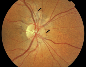
- Angioid streaks of the retina
- Blue sclerae
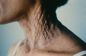
- Cutaneous laxity
- Gastrointestinal bleeding
- High arched palate
- Hypertension
- Joint hypermobility
More detailed description of symptoms
X-ray abnormalities
- Intracranial calcification
Congenital conditions
Cardiac and vascular conditions
- Mitral valve incompetence
Respiratory conditions
DIAGNOSIS
Diagnosis relies on clinical recognition of skin lesions and histologic demonstration of calcification of elastic fibers that are clumped and fragmented in the dermis. Elastic fibers altered in patients with pseudoxanthoma elastic stain blue with hematoxylin /eosin due to their calcium content.
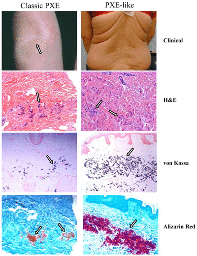
The role of connective tissue components in the etiology of pseudoxanthoma elasticum includes changes in collagen, elastin, glycosaminoglycans, fibronectin, microfibrillar proteins, modifying enzymes of extracellular matrix proteins, fibroblasts, and fibrillins.
Recognition of mutations in the ABCC6 gene provides a means for prenatal test in families that are at risk.
PATHOGENESIS /MUTATION
PXE is associated with mutations in the ABCC6 (ATP binding cassette subtype C number 6) gene. At least one ABCC6 mutation is found in about 80% of patients.
The main mutations consist of:
- missense
- nonsense
- frameshift mutations
- or large deletions.
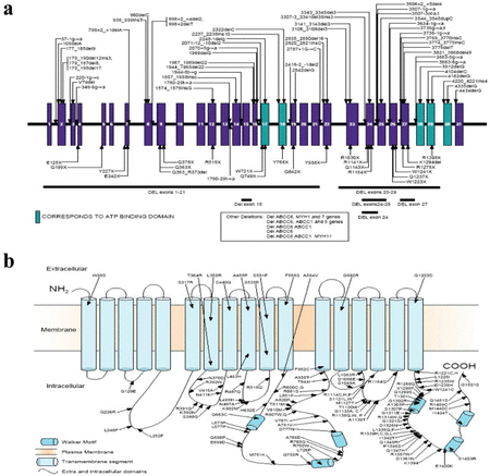
ABCC6 encodes the protein ABCC6 (also known as MRP6), a member of the large ATP dependent transmembrane transporter family that is expressed predominantly in the liver, presumably transporting critical metabolites to the circulation. In the absence of ABCC6 transporter changes in the concentration of such molecules in the circulation can take place with mineralization and fragmentation of the elastin fibers in connective tissue: Bruch's membrane in the eye, arterial blood vessel, kidney and skin.
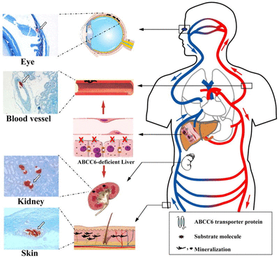
Recent studies hypothesize that PXE is a metabolic disease because metabolites of vitamin K cannot reach peripheral tissues.
PXE and vitamin k
Sometimes PXE is caused by gamma-glutamyl carboxylase mutation. It is encoded by the GGCX gene and it is dependent on reduced vitamin K as a co-factor in the liver. The oxidized forms of vitamin K and the dietary vitamin K are reduced by enzymatic reactions coupled with conversion of GSH to GSSG.
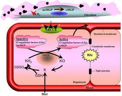
It is worthwhile mentioning that the vitamin K-dependent enzymes responsible for carboxylation of clotting factors are also involved in modulating bone mineralization and in preventing calcium precipitation in soft tissues. Sometimes mutations can occur. They are of critical importance in domains that are essential in the activating role of the GGCX enzyme in the coagulation cascade, but also for the activation of other gla-proteins such as, for example, matrix gla protein or osteocalcin. The latter are known inhibitors of calcification.
Hence, a decreased or absent activation of such proteins can explain the calcification and subsequent fragmentation of the elastic fibers. The severity of the skin lesions might be due to the additive effect of decreased or absent activity of multiple calcification inhibitors.
Vitamin k deficiency
Furthermore, apart from gene defects, the correct carboxylation of clotting factors and of modulators of mineralization in bone and soft tissues may depend on post-translational maturation of enzymes as well as on cell metabolic alterations affecting, for instance, the structural organization of membranes of the endoplasmic reticulum where carboxylating enzymes are located. Therefore, the problem is rather complex and the involvement of vitamin K-dependent processes in mineralization of connective tissues and of elastic fibers in particular is under investigation.
COMPLICATIONS
Cardiac complications and PXE may be risk factors for strokes in β-thalassemia. Their frequent coexistence leads to a therapeutic dilemma in patients requiring antithrombotic therapy.
THERAPY
- Different therapeutic strategies to treat retinal manifestations are discussed, including thermal laser coagulation, photodynamic therapy, and intravitreal injections of drugs inhibiting vascular endothelial growth factor.
- Antiangiogenic drugs such as bevacizumab and ranibizumab have been effective.
- Patients with cardiovascular disease are treated as individuals without PXE.
- Special recommendation in the use of FANS (like aspirin or ibuprofen) that increase bleeding risk.
- It is important to reduce the potential factors that contribute to the development of the disease, such as smoking and alcol.
- Skin plastic surgery can be performed routinely without complications.