Liver
Tissues

Author: Paolo Pescarmona
Date: 17/10/2011

Description

Liver zonation

Zonation of parenchymal and nonparenchymal metabolism in liver. 1996

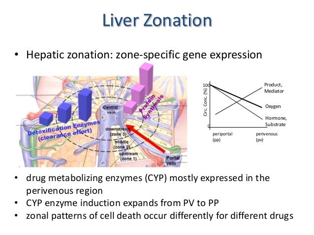
  • The enormous number of different liver functions are carried out by parenchymal and four main types of nonparenchymal cells, either alone or in cooperation. Although the liver tissue is uniform on the level of histology, it is heterogenous on the level of morphometry and histochemistry. This heterogeneity is related to the blood supply; cells located in the upstream or periportal zone differ from those in the downstream or perivenous zone in their equipment with key enzymes, translocators, receptors, and subcellular structures and therefore have different functional capacities. This is the basis of the model of metabolic zonation, according to which glucose release from glycogen and via gluconeogenesis, amino acid utilization and ammonia detoxification, protective metabolism, bile formation, and the synthesis of certain plasma proteins such as albumin and fibrinogen occur mainly in the periportal area, whereas glucose utilization, xenobiotic metabolism, and the formation of other plasma proteins such as alpha 1-antitrypsin or alpha-fetoprotein occur predominantly in the perivenous zone. The morphologic and functional heterogeneity is the result of zonal differences in the activation of the cellular genome caused by gradients in oxygen, substrate, hormone, and mediator levels, in innervation, as well as in cell-to-cell and cell-to-biomatrix interactions.

wnt hepatocytes proliferation

Perivenous expression of the mRNA of the three hypoxia-inducible factor a-subunits, HIF1a, HIF2a and HIF3a, in rat liver, 2001

Wnt signalling and the control of cellular metabolism 2010

MeSH
Comments
2013-01-19T16:46:20 - chiara magaton

Faccia un pagina

HEPATIC ENCEPHALOPATHY

Sotto Disease, ci copi questo contenuto e poi lo cancelli.

Hepatic encephalopathy (HE), also known as portosystemic encephalopathy, is a major neuropsychiatric complication of cirrhosis and it is the occurrence of confusion, altered level of consciousness, and coma as a result of liver failure. In the advanced stages it is called hepatic coma or coma hepaticum and it may ultimately lead to death.
Hepatic encephalopathy: pathophysiology and advances in therapy. 2007
HE comprises a broad range of potentially reversible neuropsychiatric disturbances in patients with liver dysfunctions after exclusion of other neurological and/or metabolic causes. For that reason the severity of hepatic encephalopathy is graded with the West Haven Criteria: this is based on the level of impairment of autonomy, changes in consciousness, intellectual function, behaviour, and the dependence on therapy.

  • Grade 1 - Trivial lack of awareness, euphoria or anxiety, shortened attention span, impaired performance of addition or subtraction;
  • Grade 2 - Lethargy or apathy, minimal disorientation for time or place, subtle personality change, inappropriate behaviour;
  • Grade 3 - Somnolence to semistupor, but responsive to verbal stimuli, confusion, gross disorientation;
  • Grade 4 - Coma (unresponsive to verbal or noxious stimuli).


Bunny .

Furthermore, a classification of hepatic encephalopathy was introduced at the World Congress of Gastroenterology 1998 in Vienna. According to this classification, hepatic encephalopathy is subdivided in type A, B and C depending on the underlying cause.
- Type A (=acute) describes hepatic encephalopathy associated with acute liver failure,
typically associated with cerebral oedema;
- Type B (=bypass) is caused by portal-systemic shunting without associated intrinsic liver
disease;
- Type C (=cirrhosis) occurs in patients with cirrhosis - this type is subdivided
in episodic,persistent and minimal encephalopathy.

Bunny .

The term minimal encephalopathy (MHE) is defined as encephalopathy that does not lead to clinically overt cognitive dysfunction, but can be demonstrated with neuropsychological studies. This is still an important finding, as minimal encephalopathy has been demonstrated to impair quality of life.
Minimal hepatic encephalopathy: time to recognise and treat. 2008

EPIDEMIOLOGY AND PROGNOSIS

In those with cirrhosis, the risk of developing hepatic encephalopathy is 20% per year, and at any time about 30–45% of people with cirrhosis exhibit evidence of overt encephalopathy. The prevalence of minimal hepatic encephalopathy detectable on formal neuropsychological testing is 60–80%; this increases the likelihood of developing overt encephalopathy in the future.
Once hepatic encephalopathy has developed, the prognosis is determined largely by other markers of liver failure, such as the levels of albumin, the prothrombin time, the presence of ascites and the level of bilirubin. Together with the severity of encephalopathy, these markers have been incorporated into the Child-Pugh score: this score determines the one- and two-year survival and may assist in a decision to offer liver transplantation.

In acute liver failure, the development of severe encephalopathy strongly predicts short-term mortality, and is almost as important as the nature of the underlying cause of the liver failure in determining the prognosis.

Bunny .

Compared to the US general population, the physical and mental component summary scores were lower in patients with cirrhosis. Among patients with cirrhosis, there were no significant differences in the physical and mental component summary scores according to age, gender, ethnicity, and aetiology. Increased severity of liver disease is associated with decreased physical aspects of quality of life. Patients with encephalopathy (overt and subclinical) had decreased physical and mental component summary scores compared to patients without encephalopathy. Compared to patients without encephalopathy, those with subclinical encephalopathy had a lower mental component summary score.
Influence of hepatic encephalopathy on health-related quality of life in patients with cirrhosis. 2003

SIGNS AND SIMPTOMS

HE develops slowly in cirrhotic patients, starting with altered sleep patterns and eventually progressing through asterixis to stupor and coma. The first stage of hepatic encephalopathy is characterised by an inverted sleep-wake pattern; the second stage is marked by lethargy and personality changes; the third is marked by worsened confusion; the fourth is marked by a progression to coma and cerebral oedema leads to death.
In the intermediate stages, a characteristic jerking movement of the limbs is observed (asterixis, "liver flap" due to its flapping character): this disappears as the somnolence worsens. There is disorientation and amnesia, and uninhibited behaviour may occur.

Flapping tremor

Increasing severity of liver disease is associated with decreased physical aspects of quality of life: overt hepatic encephalopathy negatively affects both physical and mental aspects of quality of life, whereas subclinical encephalopathy affects mainly the mental aspects, independently of age, gender, ethnicity, aetiology and liver disease severity. Widespread cortical and subcortical network connectivity changements that correlated with neuropsychologic impairment were found in patients with subclinical HE. In particular, impairment in the basal ganglia-thalamocortical circuit could play an important role in mediating neurocognitive dysfunction, especially for psychomotor speed and attention deficits in patients with subclinical HE. Additionally, studies revealed significantly reduced functional connectivity in the right middle frontal gyrus and left posterior cingulate cortex in the HE patients.
Encephalopathy often occurs together with other symptoms and signs of liver failure. These may include jaundice, ascites, and peripheral edema. The tendon reflexes may be exaggerated, and the plantar reflex may be abnormal, namely extending rather than flexing (Babinski's sign) in severe encephalopathy. A particular smell (foetor hepaticus) may be detected.

CAUSES

In a small proportion of cases, the encephalopathy is caused directly by liver failure; this is more likely in acute liver failure. More commonly, especially in chronic liver disease, hepatic encephalopathy is caused or aggravated by an additional cause, and identifying these causes can be important to treat the episode effectively. These predisposing factors are common and include:
° Excessive nitrogen load, caused by:
- Oral protein load
- Gastrointestinal bleeding
- Renal failure
° Electrolyte or metabolic disturbances:
- Hyponatraemia
- Hypokalaemia
- Diuretics abuse (generally used for the treatment of ascites)
- Hypoxia
- Dehydratation
° Drugs:
- Sedatives
- Narcotics
- Antipsychotics
- Alcohol intoxication
° Infections, especially by HCV
° Transjugular intrahepatic portosystemic stent shunts (TIPS)
° Poor nutritional status
° DM and insulin resistance
° Surgery
° Unknown: in 20–30% of cases, no clear cause for an attack can be found.

PATHOGENESIS

The pathogenesis of HE in cirrhosis is complex and multifactorial, but a key role is thought to be played by circulating gut-derived toxins of the nitrogenous compounds, most notably ammonia.
In healthy subjects, nitrogen-containing compounds from the intestine, generated by gut bacteria from food, are transported by the portal vein to the liver, where 80–90% is metabolised through the urea cycle and/or excreted immediately.

 Bunny.

This process is impaired in all subtypes of hepatic encephalopathy, either because the hepatocytes are incapable of metabolising the waste products or because portal venous blood bypasses the liver through collateral circulation or a medically constructed shunt. Nitrogenous waste products (ammonia, NH3, is the most important one) accumulate in the systemic circulation (hence the older term "portosystemic encephalopathy"). Hyperammonemia contributes to the confusion and coma of hepatic encephalopathy.
The brain is much more susceptible because ammonium crosses the blood-brain barrier and is absorbed and metabolised by the astrocytes. Astrocytes use ammonia when synthesising glutamine from glutamate. The increased levels of glutamine lead to an alteration in the expression of key astrocytic proteins and to an increase in osmotic pressure in the astrocytes, which become swollen: these changes are known as Alzheimer type II astrocytosis.

Brain-blood ammonia concentration ratios (normally of the order of 2) are increased up to fourfold in liver failure and arterial blood ammonia concentrations are good predictors of cerebral herniation in patients with acute liver failure. Functional studies of ammonia-treated astrocytes have shown the following: with low doses or short-term exposure, the uptakes of K+, glutamate and GABA remained unchanged or slightly increased, whereas with higher doses or longer treatment, those activities diminished. A fall in ATP values occurred with prolonged ammonia treatment.
The role of astrocytes in hepatic encephalopathy. 1987
Since the CNS is devoid of effective urea cycle activity, ammonia removal by brain relies on glutamine formation. Cerebrospinal fluid and brain glutamine are found to be significantly elevated in cirrhotic patients with encephalopathy and in rats following portocaval anastomosis. In both cases, glutamine is found to be elevated in a region-dependent manner. The increased formation of brain glutamine in hyperammonaemic syndromes could be responsible for the phenomenon of brain oedema in these disorders.
Glutamine synthetase in brain: effect of ammonia. 2002
Neurobiology of ammonia. 2002
Ammonia: key factor in the pathogenesis of hepatic encephalopathy. 1987
Several mechanisms have been proposed to explain the neurotoxic action of ammonia. Such mechanisms include
(Alterations in expression of genes coding for key astrocytic proteins in acute liver failure. 2001 ; Pathophysiology of hepatic encephalopathy: a new look at ammonia. 2002 ; Ammonia and proinflammatory cytokines modify expression of genes coding for astrocytic proteins implicated in brain edema in acute liver failure. 2010):
- Modification of blood-brain barrier transport with a redistribution of cerebral blood flow;
- Alterations of cerebral energy metabolism, which is redistributed from cortical to sub-cortical structures: magnetic resonance spectroscopic studies reveal increased brain lactate concentrations that are positively correlated with severity of encephalopathy and brain edema in acute liver failure, suggesting a deficit of cellular oxidative capacity and impending brain energy failure. It has been also observed a deficit in cerebral energy metabolism due to ammonia-induced inhibition of alpha-ketoglutarate dehydrogenase;
Effects of hyperammonaemia on brain function. 1998
- Direct actions on the neuronal membrane;
- Osmotic effects of an increase in astrocytic glutamine concentrations and inhibition of glutamate removal from brain extracellular space by both astrocytic and neuronal elements: hyperammonemia could induce the formation of inducible NOS in astroglial cells, with the consequent NO formation, deactivation of glutamine synthetase (GS) and dawn-regulation of glutamate uptake. Other mechanisms involved are: release due to swelling, reversal of glutamate transporters and release due to Ca2+-dependent vesicular release;
- Increase in GABA-induced chloride current in cultured neurons: probably by modifying the affinity of the GABA receptor for GABA and enhancing synergistically the binding of a GABA agonist and a benzodiazepine (BZ) agonist to the GABA receptor complex, phenomena which would enhance the neuroinhibitory effects of these ligands. In addition, ammonia has been shown to inhibit astrocytic uptake of GABA by 30%-50%, an effect which would increase the synaptic availability of GABA at GABA receptors;
- Post-translational modifications of the serotonin and noradrenaline transporters resulting in increased extracellular concentrations of these monoamines;
- Impairment of the function of the glutamate-nitric oxide-cGMP pathway in brain: the increased activation of soluble guanylate cyclase by nitric oxide (NO) in lymphocytes and in cerebral cortex is responsible for reduced ability to learn some types of tasks and so restoring the pathway and cGMP levels in brain restores learning ability
Cyclic GMP pathways in hepatic encephalopathy. Neurological and therapeutic implications. 2010 ;
- Alterations in the expression of genes coding for key astrocytic proteins, including:
° The structural glial fibrillary acidic protein (GFAP);
° Glutamate transporters GLT-1 and GLAST: this results in reduced high affinity uptake sites for glutamate, in a reduction in glutamate transport sites in brain and in a consequent increase in extracellular brain glutamate concentrations, which may be responsible for the hyperexcitability and the cerebral oedema observed in hyperammonaemic disorders;
Glutamate transporters in hyperammonemia. 2002
° Peripheral-type (mitochondrial) benzodiazepine receptors (PTBR);
° Glucose transporter (GLUT-1);
° The water channel aquaporin IV: astrocytes modulate AQP4 mRNA expression and distribution and the aggregation status of the ensuing protein depending on ammonia concentration and duration of exposure, determining a smaller water influx in ammonia-treated astrocytes. Furthermore, AQP4 gene suppression determines the appearance of a new morphological cell phenotype associated with a strong reduction in cell growth.
Ammonia induces aquaporin-4 rearrangement in the plasma membrane of cultured astrocytes. 2012

Despite numerous studies demonstrating the central role of ammonia, ammonia levels don't always correlate with the severity of the encephalopathy. Besides, inflammatory cytokines and reactive oxygen species appear to play a crucial role in the pathogenesis of HE.
There is evidence to suggest that, in acute liver failure (ALF), brain ammonia and proinflammatory cytokines may act synergistically to cause brain oedema and its complications (intracranial hypertension, brain herniation). It seems that ammonia and proinflammatory cytokines modify the expression of genes coding for astrocytic proteins (AQP4, GFAP, iNOS, HO-1) and, although modest, these effects are additive, suggesting a synergistic mechanism. However, the molecular mechanisms involved remain to be established.
Additionally, recent studies indicate that other suspected toxins in HE, such as phenol, produce comparable effects to that caused by ammonia, particularly on the astrocyte benzodiazepine receptor.

DIAGNOSIS

In general, it is based on clinical findings, whereas measurement of serum ammonia has no validated role in HE diagnosis.
The diagnosis is usually made by neuropsychological and/or neurophysiological testing in cirrhotic patients who are otherwise normal on neurological examination. At present the most frequently used psychometric methods for diagnosing minimal hepatic encephalopathy are the Inhibitory Control Test and the Psychometric Hepatic Encephalopathy Score (PHES). Another frequently used method is Critical Flicker Frequency. Today, no data are available that allow to decide which of these methods is the most appropriate. Visually evoked potentials (P300 wave) and critical flicker frequency analysis are also objective and sensitive techniques for detection of minimal HE.
It is recommended to screen all patients with cirrhosis for minimal hepatic encephalopathy using psychometric testing because it impairs the health-related quality of life, predicts the development of overt encephalopathy and is probably associated with a poor prognosis.
[Diagnostics and treatment of hepatic encephalopathy]. 2012 ; Psychometric tests for diagnosing minimal hepatic encephalopathy. 2012

Additionally, cerebral magnetic resonance imaging reveals that marked loss of brain tissue density is common in cirrhosis, progresses during the course of the disease, is greater in patients with history of hepatic encephalopathy and persists after liver transplantation.
Cerebral magnetic resonance imaging reveals marked abnormalities of brain tissue density in patients with cirrhosis without overt hepatic encephalopathy. 2011 ; Altered brain functional connectivity in patients with cirrhosis and minimal hepatic encephalopathy: a functional MR imaging study. 2012 ; Altered resting-state brain activity at functional MR imaging during the progression of hepatic encephalopathy. 2012

PREVENTION AND TREATMENT

Prevention and treatment of HE in cirrhotic patients continue to rely on ammonia-lowering strategies which include:
- Assessment of dietary protein intake;
- Use of non-absorbable disaccharides, such as Lactulose: they have traditionally been regarded as first-line pharmacotherapy for patients with HE, but multiple adverse events have been associated with their use;
- Use of antimicrobials, such as Neomycin, Metronidazole, Vancomycin and Rifaximin: they may be used as alternative treatments for patients intolerant or unresponsive to non-absorbable disaccharides and they reduce bacterial production of ammonia and other bacteria-derived toxins through suppression of intestinal flora;
- Use of Sodium benzoate;
- Use of Mannitol;
- Use of L-ornithine- L-aspartate LOLA: it reduces blood ammonia concentrations and brain water content significantly and results in a significant delay in onset of severe encephalopathy
A randomized controlled trial comparing lactulose, probiotics, and L-ornithine L-aspartate in treatment of minimal hepatic encephalopathy. 2011 ;
- Use of the benzodiazepine receptor antagonist Flumazenil;
- Use of dopamine receptor agonists;
- Use of probiotics;
- Use of inhibitors of phosphodiesterase 5 (PDE-5), such as Zaprinast and Sildenafil, or anti-inflammatories, such as Ibuprofen: they increase cGMP levels
Mechanisms of cognitive alterations in hyperammonemia and hepatic encephalopathy: therapeutical implications. 2009 ;
- Mild hypothermia: it delays in the onset of severe encephalopathy, selectively normalizes lactate and alanine synthesis from glucose, prevents the impairment of oxidative metabolism and results in a reduction of brain water content, in a significant reduction of cerebrospinal fluid (CSF), but not plasma ammonia concentrations and in an inhibition of blood-brain transfer of ammonia. It seems to offer a potentially useful bridge therapy in patients with acute liver failure who are awaiting liver transplantation. The impact of potential adverse events, such as infection, should also be taken into account
Mild hypothermia for acute liver failure: a review of mechanisms of action. 2005 ; Therapeutic hypothermia for acute liver failure. 2009

Instead, strategies aimed at lowering gut ammonia production are generally ineffective.

Pharmacotherapy for hepatic encephalopathy. 2010

AddThis Social Bookmark Button