Epilepsy
Diseases

Author: Gianpiero Pescarmona
Date: 28/02/2010

Description

DEFINITION

Epilepsy (from the Ancient Greek ἐπιληψία (epilēpsía) — "to seize") is a common chronic neurological disorder characterized by recurrent unprovoked seizures

DatabaseLink
The Diseases DatabaseURL

Epilepsy

EPIDEMIOLOGY

age, sex, seasonality, etc

SYMPTOMS

DIAGNOSIS

histopathology
radiology
NMR
laboratory tests

PATHOGENESIS

As a working hypothesis we can assume a major role of GABAergic system as a causal factor of epilepsy. The affected step can be:

glycyrrhetinic acid epilepsy

PATIENT RISK FACTORS

Epilessia, studio Telethon inchioda gli 'aiutanti' dei neuroni

Roma, 14 mag. (Adnkronos Salute) - L'epilessia non si può imputare soltanto ai neuroni: anche altre cellule del cervello, come gli astrociti, hanno un ruolo importante nella genesi delle scariche elettriche tipiche di questa malattia neurologica. Lo dimostra uno studio finanziato da Telethon e dalla Commissione europea, pubblicato sulle pagine di 'Plos Biology'. Il lavoro è il risultato di una collaborazione tra tre diversi gruppi di ricerca legati all'Istituto di neuroscienze del Cnr di Padova e Pisa e all'Istituto neurologico Besta di Milano, coordinati da Giorgio Carmignoto, Gian Michele Ratto e Marco de Curtis. I ricercatori sono riusciti a dimostrare il ruolo attivo nella nascita delle scariche epilettiche degli astrociti, cellule gliali e non neuronali. Sono molto numerose nel cervello dei mammiferi, e dialogano continuamente con i neuroni. Lo studio dimostra come l'interazione tra neuroni e astrociti sia uno dei meccanismi che contribuisce alla generazione delle scariche epilettiche. Ritenuti in passato dei semplici 'aiutanti' dei neuroni, gli astrociti si sono rivelati nel corso del tempo cellule che esercitano nel cervello un ruolo decisamente più attivo. Questo vale evidentemente anche per la genesi delle crisi epilettiche. Monitorando in laboratorio l'attività di neuroni e astrociti, in diversi modelli sperimentali, i ricercatori hanno infatti scoperto che nella zona di generazione delle scariche epilettiche gli astrociti sono in grado di amplificare nei neuroni circostanti lo stato di ipereccitabilità, che può poi tradursi nella scarica epilettica. A riprova di questo, i ricercatori hanno constatato che inibendo l'attività degli astrociti si riducono le scariche epilettiche, e viceversa. Per chi studia l'epilessia, chiarire i meccanismi biologici, tuttora ben poco conosciuti, che portano allo scatenarsi delle crisi epilettiche è fondamentale per poter sviluppare cure. L'epilessia è una patologia cerebrale che può avere basi genetiche oppure essere la conseguenza di malformazioni del cervello, traumi, infezioni, ictus o tumori. Altre volte la causa è addirittura sconosciuta. Qualsiasi sia l'origine, la manifestazione tipica della patologia epilettica è rappresentata sempre da crisi convulsive, più o meno frequenti, che se non opportunamente trattate possono mettere a rischio la sopravvivenza del paziente. Queste crisi sono la conseguenza di un'anomalia nell'attività elettrica dei neuroni, che raggiungono una sorta di ipereccitazione diffusa ed esageratamente sincrona. Ad oggi non esiste una cura risolutiva: l'unico trattamento disponibile è a base di farmaci capaci di arrestare le convulsioni, ma non di eliminare i meccanismi anomali che sono la causa dell'equilibrio alterato del tessuto nervoso. Inoltre, questi farmaci sono inefficaci in circa un terzo dei pazienti che tendono a sviluppare un'epilessia cronica, spesso accompagnata da gravi problemi neurologici e relazionali.Questo studio rappresenta dunque un significativo passo in avanti nella comprensione dei meccanismi cellulari alla base della patologia epilettica e potrebbe aiutare a delineare una nuova strategia terapeutica per l'epilessia che abbia nell'attività degli astrociti il bersaglio principale.

Genetic

Debate: Does genetic information in humans help us treat patients? PRO--genetic information in humans helps us treat patients. CON--genetic information does not help at all. 2008
Epilepsia. 2008 Dec;49 Suppl 9:13-24.
Delgado-Escueta AV, Bourgeois BF.

  • PRO: In the past decade, genotyping has started to help the neurologic practitioner treat patients with three types of epilepsy causing mutations, namely (1) SCN1A, a sodium channel gene mutated in Dravet's sporadic severe myoclonic epilepsy of infancy (SMEI and SMEB); (2) laforin (dual specificity protein phosphatase) and malin (ubiquitin E3 ligase) in Lafora progressive myoclonic epilepsy (PME); and (3) cystatin B in Unverricht-Lundborg type of PME. Laforin, malin, and cystatin B are non-ion channel gene mutations that cause PME. Genotyping ensures accurate diagnosis, helps treatment and genetic counseling, psychological and social help for patients and families, and directs families to organizations devoted to finding cures for specific epilepsy diseases. In SCN1A and cystatin B mutations, treatment with sodium channel blockers (phenytoin, carbamazepine, oxcarbazepine, lamotrigine) should be avoided. Because of early and correct diagnosis by genotyping of SCN1A mutations, the avoidance of sodium channel blockers, and aggressive treatment of prolonged convulsive status, there is hope that Dravet's syndrome may not be as severe as observed in all past reports. Genotyping also identifies nonsense mutations in Lafora PME. Nonsense mutations can be corrected by premature stop codon readthrough drugs such as gentamicin. The community practitioner together with epilepsy specialists in PME can work together and acquire gentamicin (Barton-Davis et al., 1999) for "compassionate use" in Lafora PME, a generalized lysosome multiorgan storage disorder that is invariably fatal. In Unverricht-Lundborg PME, new cohorts with genotyped cystatin B mutations have led to the chronic use of antioxidant N-acetylcysteine and combination valproate clobazam or clonazepam plus antimyoclonic drugs topiramate, zonisamide, piracetam, levetiracetam, or brivaracetam. These cohorts have minimal ataxia and no dementia, questioning whether the syndrome is truly progressive. In conclusion, not only is genotyping a prerequisite in the diagnosis of Dravet's syndrome and the progressive myoclonus epilepsies, but it also helps us choose the correct antiepileptic drugs to treat seizures in Dravet's syndrome and Unverricht-Lundborg PME. Genotyping also portends a brighter future, helping us to reassess the true course, severity, and progressive nature of Dravet's syndrome and Unverricht-Lundborg PME and helping us craft a future curative treatment for Dravet's syndrome and Lafora disease. Without the genotyping diagnosis of epilepsy causing mutations we are stuck with imprecise diagnosis and symptomatic treatment of seizures.
  • CON: Genotyping of epilepsy may help to better understand the genetics of epilepsy, to establish an etiology in a patient with epilepsy, to provide genetic counseling, and to confirm a clinical diagnosis. However, critical analysis reveals that genotyping does not contribute to an improved treatment for the patients. In order to improve treatment, genotyping would have to (1) improve our ability to select the drug of choice for a given epilepsy or epileptic syndrome; (2) improve our ability to predict the individual risk of adverse reactions to certain drugs; (3) improve our ability to avoid unnecessary treatments or treatments that could aggravate seizures. Many example illustrate the lack of impact of genetic information on the treatment outcome: we do not treat Dravet syndrome more successfully since SCN1A testing became available; we do not treat Lafora disease more successfully since testing for laforin and malin became available; we do not need to know the genetic nature of Unverricht-Lundborg disease or test for the cystatin B mutation in order to select or avoid certain drugs; we do not treat Rett syndrome more successfully since MECP2 testing became available; we do not treat JME more successfully since we know its genetic origin; we do not treat autosomal dominant nocturnal frontal lobe epilepsy more successfully since we know its genetic origin and can test for its mutation. The clinical characteristics as well as the response to treatment of these epilepsy syndromes have been well established before genotyping became available. It can not be argued that genotyping is necessary for establishing a diagnosis or ensure accurate diagnosis. Since not all individuals with given syndromes have been shown to have the corresponding mutation, the clinical diagnosis must have been based on well-established clinical criteria. In addition, the presence or absence of the mutation in a given patient has never been shown to specifically predict the response to any form of treatment, positive or negative. Finally, the appropriate psychological and social help in a given patient will not depend on the identification of a mutation. This does not leave any role for genotyping in epilepsy for the sole reason of improving treatment of the patient. Claiming that the result of genotyping predicts optimal treatment in certain epilepsies is equivalent to stating that genotyping for diabetes has become available and that, based on this breakthrough, insulin can now be selected as the treatment of choice in those who test positive.

Vascular

Genetic

Acquired

Hormonal

Genetic

Acquired

TISSUE SPECIFIC RISK FACTORS

anatomical (due its structure)

vascular (due to the local circulation)

physiopathological (due to tissue function and activity)

COMPLICATIONS

THERAPY

  • increasing GABA production

ABSTRACT (The ketogenic diet (KD) is a high-fat, low-carbohydrate, and adequate-protein diet used to treat drug-resistant seizures) 2005

Kirk Nylen, Sergei Likhodii, Peter A. Abdelmalik, Jasper Clarke, and W. McIntyre Burnham

The ketogenic diet (KD) is a high-fat, low-carbohydrate, and adequate-protein diet used to treat drug-resistant seizures. It is currently unknown exactly how the KD exerts its anticonvulsant effects. Animal models are used to study the KD anticonvulsant mechanism of action. In the present study, we explored the ability of two KDs, a 4:1 KD and a 6.3:1 KD (more severe ketosis), to elevate pentylenetetrazole (PTZ) seizure thresholds in adult rats and young rats. When calculating PTZ thresholds, one can use either "absolute latencies" (i.e., the number of seconds until a seizure occurs) or "threshold doses" (i.e., the milligram/kilogram dose of PTZ required to elicit a seizure). When absolute latencies were used, neither KD significantly increased the latency to seizure in adult rats and young rats. Similarly, neither KD elevated threshold doses in adult rats. The 4:1 KD did not elevate threshold doses in young rats; however, the 6.3:1 KD did. Threshold doses are sensitive to differences between group weights, and the largest difference between group weights existed between the 6.3:1 KD group and its respective control group. It remains unclear whether threshold doses, absolute latencies, or some combination of the two methods should be used when determining seizure thresholds in rats fed a KD and seizure tested by using the PTZ infusion test. We conclude that the PTZ-infusion test is not suitable for modeling the anticonvulsant effects of the KD seen clinically, especially when dietary treatments lead to significantly mismatched body weights between the groups.

Neurosteroids Analogs

Neurosteroids and epilepsy. 2010
Curr Opin Neurol. 2010 Apr;23(2):170-6.
Biagini G, Panuccio G, Avoli M.

Dipartimento di Scienze Biomediche, Università di Modena and Reggio Emilia, Modena, Italy.
Abstract

PURPOSE OF REVIEW: Neurosteroids are a family of compounds synthesized directly in the brain by transforming cholesterol into pregnenolone, which is then converted to compounds such as allopregnanolone and allotetrahydrodeoxycorticosterone. In view of their ability to modulate neurotransmission, neurosteroids may influence the clinical course of epileptic disorders. In this review, we highlight two emerging properties of neurosteroids, that is, their anticonvulsant and antiepileptogenic activities. RECENT FINDINGS: It has been shown that fluctuations in neurosteroid synthesis, such as those seen in response to stress or during the ovarian cycle, determine an increase in seizure threshold. Moreover, increased neurosteroid synthesis, presumably occurring in glial cells during epileptogenesis, delays the appearance of recurrent spontaneous seizures in an animal model of temporal lobe epilepsy; such an effect may be due to augmented tonic gamma-aminobutyric acid type A receptor-mediated inhibition. Finally, clinical trials with ganaxolone, an allopregnanolone analogue, have demonstrated beneficial effects in pharmacoresistant epileptic patients, whereas finasteride--which interferes with neurosteroid synthesis - facilitates seizures in catamenial epilepsy. SUMMARY: The overall evidence suggests that neurosteroids may represent a novel therapeutic strategy in epileptic disorders and a future perspective to control epileptogenicity.

Update on the Neurobiology of Alcohol Withdrawal Seizures, 2005

  • Abrupt cessation of alcohol intake after prolonged heavy drinking may trigger alcohol withdrawal seizures. Generalized tonic–clonic seizures are the most characteristic and severe type of seizure that occur in this setting. Generalized seizures also occur in rodent models of alcohol withdrawal. In these models, the withdrawal seizures are triggered by neuronal networks in the brainstem, including the inferior colliculus; similar brainstem mechanisms may contribute to alcohol withdrawal seizures in humans. Alcohol causes intoxication through effects on diverse ion channels and neurotransmitter receptors, including GABAA receptors—particularly those containing δ subunits that are localized extrasynaptically and mediate tonic inhibition—and N-methyl-D-aspartate (NMDA) receptors. Alcohol dependence results from compensatory changes during prolonged alcohol exposure, including internalization of GABAA receptors, which allows adaptation to these effects. Withdrawal seizures are believed to reflect unmasking of these changes and may also involve specific withdrawal-induced cellular events, such as rapid increases in α4 subunit–containing GABAA receptors that confer reduced inhibitory function. Optimizing approaches to the prevention of alcohol withdrawal seizures requires an understanding of the distinct neurobiologic mechanisms that underlie these seizures.

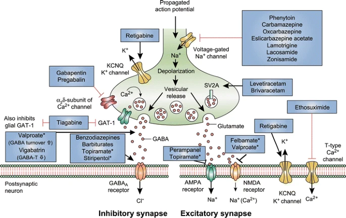
Mechanism of action of clinically approved anti-seizure drugs. Updated and modified from Löscher and Schmidt [151]. Drugs marked with asterisks indicate that these compounds act by multiple mechanims (not all mechanisms shown here). GABA-T GABA aminotransferase, GAT GABA transporter, SV2A synaptic vesicle protein 2A, GABA gamma-aminobutyric acid, NMDA N-methyl-D-aspartate, AMPA α-amino-3-hydroxy-5-methyl-4-isoxazolepropionic acid, KCNQ a family of voltage-gated potassium channels (also known as the Kv7 family)

Synaptic Vesicle Glycoprotein 2A Ligands in the Treatment of Epilepsy and Beyond, 2018

Brivaracetam: Rationale for discovery and preclinical profile of a selective SV2A ligand for epilepsy treatment, 2016

epilepsy and statin

epilepsy+and+anandamide

Comments
2021-05-25T15:39:09 - Gianpiero Pescarmona

Alternative Approaches to Therapy

Increasing ATP production
Physiological Channels Agonists/Antagonists

2014-06-15T13:47:05 - Virginia Eterno

Overview of Epilepsy

Epilepsy is one of the most common disorders of the brain, known since antiquity. It affect an exstimated 50 million people worldwide, of all ages.
This disorders seriously compromises quality of life, and it is also associated with social disadvantages, and results in increased morbidity and premature mortality.

The modern neurobiological analysis of epilepsy began with John Hughlings Jackson’s work, in the 1860s.
The International League against Epilepsy (ILAE) proposed the following definition for epileptic seizures and epilepsy: “ Epilepsy is a chronic disorder of the brain characterized by an enduring predisposition to generate epileptic seizures and by the neurobiological, cognitive, psychological and social consequences of this conditions . The definition of Epilepsy requires the occurrence of at least one epileptic seizure ”.

Seizures are temporary disruptions of brain function resulting from abnormal, excessive or hypersynchronous neuronal activity. (Kandel et al, Principles of Neural Science, McGraw-Hill, 5th edition)
Epileptic seizures are characterized by brief episodes of loss of awareness (absence) and sensitive, psychic and motor alterations with skeletal muscle contractions.
The ILAE developed an international classification of epileptic seizures that separates seizures into two categories:

  • Focal or partial seizures: originate in a small group of neurons (seizure focus) and the symptoms depend on the location of the focus within the brain is often preceded by auras, that is caused by electrical activity originating from seizure focus. Focal seizures can be very brief or last for minutes. Sometimes, epileptic activity starts as a focal seizure, spreads to the rest of the brain and becomes a generalized seizure.
  • Generalized seizures: involve both hemispheres and it can be classified in a convulsive type or non-convulsive type, depending on whether the seizures is associated with tonic or clonic movements.
    The distinctive clinical patterns of focal seizures and generalized seizures can be attribute to the different patterns of activity of cortical, subcortical or spinal cord neurons.

The EEG (Electroencephalograph)is a measure of the electrical activity of the brain, which dramatically increases during a seizure or immediately after; (Zhang et al, Transition to seizure: From Macro-to-Micro mysteries, Epilepsy Research, 2011) this huge rise of electrical activity of the cells is detectable in the EEG trace, which becomes similar to the trace of a seismograph during an earthquake. Here is depicted an example of a EEG traces recorded from a patient during a seizure and one from an healthy individual.

Electrophysiology

The enhanced excitability (epileptiform activity) may result from many different factors, such as altered cellular properties or altered synaptic connections. (Beck and Yaary, Plasticity of intrisic neuronal properties in CNS disorders. Nature Review Neuroscience, 2008) Each neuron within a seizure focus has a stereotypic and synchronized electrical response called paroxysmal depolarizing shift (PDS) (Altrup, Epileptogenicity and epileptic activity: mechanisms in an invertebrate model nervous system, Curr Drug Target, 2004), an intracellular depolarization that is sudden, large (20-40 mV) and long lasting (50-200 ms), triggers a train of action potentials and is followed by an after-hyperpolarization that is generated by several types of K+ channels and limits the duration of the PDS. The neurons involved fire all together and lose their correct functions: PDS is associated with alterations in spike generation, decrease in amplitude, increase in spike duration and decrease in the slope of rise and decay.

Pathogenesis

Despite the high prevalence of this neurological disorders and the extensive research on it, the cellular and molecular mechanism, underlying epileptogenesis still remain unclear. Epilepsy is in the majority of cases multifactorial and is the result of genetic and acquired influences and provoking factors.

We know that the gradual loss of GABAergic inhibition is critical to the early steps in this progression, together with a chronic modifications in dendrite morphology, membrane channels or extracellular ions, but the factor leading to development of epilepsy are un unfolding mistery.
But also an head trauma, strokes, brain tumors, alcohol or drug, damage of the blood brain barrier (Seiffert et al, Lasting blood-brain barrier disruption induces epileptic focus in the rat somatosensory cortex, Journal of Neuroscience, 2004), alteration of the extracellular matrix, defects on the correct development of the network, such as angiogenesis or synaptic plasticity, and even axonal and myelin injury, neurodegeneration, channelopaties, gliosis, neurogenesis, inflammatory processes have the functional outcome to the epileptic disease. (Kharatishvili and Pitkanen, Posttraumatic epilepsy, Current opinion in Neurology, 2010)

Genetic

Moreover, some form of epilepsy are caused in part by a genetic predisposition: at present, we know 1000 human genes have been linked to an epileptic phenotype and, among these, about 70 genes codify for ion channels. The affected proteins include also protein involved in synaptic transmission such as transporters, vesicle proteins, synaptic receptors and molecules involved in Ca2+ signalling. (Noebels, The biology of epilepsy genes. Annu Rev Neuroscience, 2003)

We can classified into three functional classes the mutations that leads to an inherited epilepsy:

1) Defects that correlate to plasmatic membrane and synaptic signalling ( channelopaties ):

Multiple examples of ion channel mutations linked to paroxysmal network synchronization have been identified, including the pore-forming and regulatory subunits of Na+, K+, Ca2+ channels, along with channels gated externally by GABA, glutamate, Ach, 5-HT (serotonin) and neuropeptide Y (NPY).

  • Voltage gate Na + channels: mutations in up to three on thirteen genes codifying for these channel subunits (Nav 1.1 in dendrites, 1.2 in axonal membranes, beta1, widely expressed). They are responsible for an inapt activation of the sodium current with following slow membrane depolarization and repolarization defects due to an incomplete inactivation of the channel.
  • Voltage gated K+ channels. Generally the channelopaties associated to these channels lead to a lasting depolarizing phase of the neuronal membrane. Different mutations within the same subunit lead to diverse clinical phenotypes, for example, human mutation of KCNA1 differentially affect assembly, targeting and kinetics of the channel and may lead to partial lobe epilepsy (if the K+ currents are severely reduced), episodic ataxia, neuromyotonia (if the current amplitude is not altered) or myokymia.
  • Voltage gated Ca 2+ channels. Mutation in up to four on twentyfour genes codifying for Ca 2+ channel were found. They are supposed to lead mainly to an impairment of the inward depolarizing current.
    This observations underlie that in presence of such mutations the ion currents through the neuronal plasmatic membrane are not balanced anymore and the electric misregulation leads up to an iperexcitability of the cerebral cells leading to the epileptic seizure occurring.

2) Mutations linked to neuronal metabolism and plasticity.

Recently mutations in gene codifying for proteins involved in neurotransmitter release were discovered, for example genes that belong to the large protein families that mediate the vesicle trafficking and exocytosis (35 SNARES, 60 Rabs, and 53-coat complex subunits in the human genome). Mainly are those codifying for the family proteins of Synapsin (Syn, in depth in this page). In animal models lacking these genes a clean decrease in the synaptic vesicles was found and these genes may be responsible for an imbalance of the neuronal excitability. Another gene that if mutated give epileptic phenotype is that codifyng for the vesicle protein Sv2A involved in readily releasable pool (RRP) mobility regulation. Another examples of mutations implicated are AP3delta (that lead to a loss of vesicular zinc sequestration, severe EEG hypersynchronization and seizures) e Znt3 (that leads to an increase in seizure susceptibility to kainite, but not to other convulsants), both genes are related also with autism.

The main target of epileptogenesis is the GABAergic transmission which is altered in different causes, such as the presynaptic reduction of GABA levels both for synthesis and release problems and the altered GABA receptor function. (Macdonald et al, Mutations linked to generalized epilepsy in humans reduce GABA receptor current. Exp Neurol, 2003). The effect of agonists and antagonists of GABA are been associated on the control of seizures activity; in fact, GABA-A receptors antagonists induce epileptic seizures, whereas agonists are frequently used as antiepilectic drugs.

Among the genes involved in GABA synthesis, mutation were found on the gene codifying for GAD65, which is an enzyme directly involved in the neurotransmitter synthesis and the gene EAAT1 codifying the glial transporter for the glutamate re-uptake.
The other target for epileptogenesis is the glutamate transmission and in particular the AMPA glutamate receptor seems to undergo a gain of function because of an altered editing of the pre-mRNA codifying for one of its subunits (GluRB).

3) Genetic causes linked to neuronal network development and differenziation.

Cortical dysplasias resulting from aberrant patterns of brain development are a frequent substrate for inherited seizure syndromes. The malformations may be microscopic or visible by magnetic resonance imaging and originate from diverse signalling defects affecting migration, proliferation, differentiation, and segmentation. Mutations in genes codifyng for transcription factors and protein related to the development and growth of the central nervous system (CNS) were found, for example, NeuroD/beta2, that blocks postnatal proliferation of granule cells in the dentate gyrus, when is lost, or Citron-kinase, a Rho effector regulating cytokinesis. Also the gene codifying for the tumor suppressor PTEN encoded a lipid phosphatase, if mutated, leads to neuronal overgrowth in cortex in patients with temporal lobe epilepsy.
Therefore, also epigenetic factors may contribute to epilepsy, for instance, mutations on MECP2, that is a gene that represses the genetic expression linking the histone deacetilase enzyme and is responsible, in humans, for X-linked Rett syndrome. (Noebels, 2003)

Idiopathic epilepsies are often considered to be “genetic”, even if a genetic cause is also not demonstrable. It seems very likely that the genetic influences in idiopathic epilepsies probably are complex involving multiple genes and interactions between genes (epistatic) and between genes and the environment (epigenetic). (Shorvon, The concept of symptomatic epilepsy and the complexities of assigning cause in epilepsy, Epilepsy Behavior, 2014).

Treatment

Although current antiepileptic medication effectively controls seizures in approximately 70% of people receiving optimal care, these medications are inadeguate for the remaining 30% of patients. (Perry and Duchowny, Surgical versus medical treatment for refractory epilepsy: outcomes beyond seizure control, Epilepsia, 2013). Medical intractability may be predicted after failure of two antiepileptic drugs.
Fewer than 10% of patients with drug refractory epilepsy are considered for surgical resection, leaving many epilepsy sufferers with no therapeutic recourse. On one hand, there is a great need for new active drugs; on the other hand, to develop such treatments, a better understanding of newly tested substances in animal models of epilepsy is required. (Loscher, Critical review of current animal model of seizures and epilepsy used in the discovery and development of new antiepileptic drugs, Seizure, 2011) Limited progress has been made since the early 1990s in the development of antiepileptic drugs with improved efficacy or tolerability. Moreover, the number of patients with drug-resistant epilepsies has not decreased, providing an impetus for development of the new, more effective antiepileptic drug treatments. In 2012, out of 30,000 compounds screened by the Anticonvulsant Screening Program (ASP) of the National Institute of Neurological Disorders and Stroke (NINDS), only 9 have acquired an indication for seizures treatment.

The molecular and cellular data on processes that underlie epileptogenesis suggest a wide spectrum of treatment targets.

  • Antiepileptic drugs (AEDs) are anticonvulsants. Commercially there are around 26 and are used to treat seizures. Here it is explained what the different AEDs are, what type of seizures or epilepsy they are used for, as well as some essential information about average doses and common side effects.
  • Surgery is an established option for those with lesion-associated epilepsy (neoplastic lesions, vascular malformations) or certain types of temporal lobe epilepsy (TLE).
  • Other novel treatment approaches for refractory patients are gene therapy drugs, including focal drug delivery and stem cell grafting. Gene therapy has significantly advanced in both preclinical and clinical research venues for the study and treatment of epilepsy. The predominant therapeutic strategy has focused upon attenuating the seizures through manipulation of excitatory or inhibitory function in the CNS. So, targets have included excitatory neurotransmitter receptors or inhibitory GABA receptors, but also the neuropeptide Y, the galanine and possibly somatostatin viral vector-based expression can result in seizure attenuation or prevention. Moreover, were also been studied the expression of the glial derived neurotrophic factor (GDNF) that protect brain cells from epilepsy damage, and the endogenous anticonvulsant, adenosine (ADK), that suppress seizure activity in brain and it is downregulated in seizure foci of epileptic patients. (Weinberg and McCown, Current prospects andchallenges for epilepsy gene therapy, Exp Neurol, 2013)
  • Vagus nerve stimulation (VNS) is a treatment for epilepsy where a small device is implanted under the skin below the left collar bone. This device, similar to a pace-maker, is called a generator. The generator is connected to a thin wire, which stimulates the vagus nerve in the person’s neck at regular times throughout the day. This sends impulses to the brain, which helps to prevent electrical activity that causes seizures. (Banerjee and Das, Refractory epilepsy, J Assoc Physicians India, 2013.)
  • Deep brain stimulation delivers electrical impulses to specific brain areas to restore the balance of circuits that are disrupted, overcoming abnormal activity in that region. DBS may be a surgical option for patients who are refractory from standard therapies.
  • Ketogenic diet (already detailed in this page)
AddThis Social Bookmark Button