Oral Candidiasis, Oral Thrush
Mycoses

Author: chiara paparella
Date: 11/01/2011

Description

Mycosis

Mycosis are infections caused by microscopic fungus. Depending on their morphology, their are divided in:

  • yeast;
  • dimorphic mycete;
  • mold

Some of thiese microorganisms are commonly being on the skin or in the organism without leading to any damage; these lead to mycosis:

  • cutaneous or mucocutaneous;
  • deep;

The first, less serious and more frequent, strikes skin, skin folds, toes, scalp, nails, mouth and genitalia. This includes candidiasi (white mouth, balanitis) and dermatophytosis (tinea, athlete foot,herpes). Sometimes it occurs if extensive antibiotics; its appearance is favourite by poor hygiene.
The second is, contrarily, a more serious form and appears only in immunosuppressed individuals (patients undergoing organ transplantation, treated with chemioterapic, immunosuppressive and corticosteroids, AIDS patients and heroin addicts). Candidias infections can take, in fact, the form of a septicemia with extension to endocardium, lungs, meninges and kidney.


Candida Albicans

The most frequent between human pathogen Candida species. This yeast occurs naturally in mouth, in gastrointestinal tract and genitalia. It usually remains confined, but, in anomalous conditions, it can be located elsewhere.
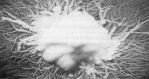
It can release about 80 different type of toxins, which can be released into circulation. After a silent period of growth, C. can evolve in fungus. It is a microorganism with a strong adaptability to environment, which it reproduce in and it can choose to differentiate into 2 forms emitting root offshoots, Rizoides.
Fungus can penetrate actively in epithelium with esoenzymes help, where cell membrane enzymes lyse host cell. It comes in the form of spores and hyphoes. The second form is regulated as one capable to invide tissues colonized and injury. This injuries are the result of the complex interaction between fungus pathogenicity, host immunitary state and organism microenvironment. The mechanisms underlying the pathogenicity are adherence to ephitelial cell, the transformation in hyohoes, cheratinolytic enzymes production (protease), virulent phenotypes selection.

Candidiasis

Candidiasis, also called Monilliasis, is a very common fungal infection.Oral candidiasis is often observed as a local opportunistic infection and is clinically divided into three types: acute forms, chronic forms, and Candida-associated lesions. Additionally, it is divided into

  • Systemic candidias;
  • Localised candidias.

The first takes place in immunocompromised individuals; it can involve the esophagus and become systemic. In severe cases C. multiplies in an abnormal manner and, through the intestine, can reach the blood where free its toxins causing Candidemia.
The second takes place in patients without impairment of immune functions and moist part of body, for ex:

  • Oral cavity (white thrush)
  • Vagina (vulvovaginitis by C.)
  • Penis (male genitalia in general)
    In cases of sistemic candidias, the disease results in bloating, slow digestion, intestinal constipation and diarrhea. Consequently there is poor absorption of nutrients.
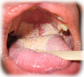
Oral Candidiasis

Strictly speaking, thrush is only a temporary candida infection in the oral cavity of babies. But we have, for this purpose, expanded the term to include candida infections occurring in the mouth and throat of adults, also known as candidiasis or moniliasis.This pathology is a fungal infection of mucosas, often this is in conjuinction with a health general compromise. In many case is caused by Candida Albicans, also called Monilia, or by Aspergillus and appear mainly in children and elderly diabetics, in wearers of dentures and in Aids patients. It can be spread to nearby organs or by blood, causing mycotic pneumonia, mycotic encephalitis, mycotic sepsis.

Symptoms

Initially, you may not have noticeable oral thrush symptoms. Signs and symptoms may develop suddenly, but they may persist for a long time and can include:

  • Creamy, white lesions on your tongue, inner cheeks and sometimes on the roof of your mouth, gums and tonsils
  • Lesions with a cottage cheese-like appearance
  • Pain
  • Slight bleeding if the lesions are rubbed or scraped
  • Cracking at the corners of your mouth
  • A cottony feeling in your mouth
  • Loss of taste

Candida overgrowth in the oral cavity may disseminate to distant organs. Therefore, in order to avoid the sequelae of systemic candidiasis, oral candidias can be rapidly controlled.

Infants and breast-feeding mothers
In addition to the distinctive white mouth lesions, infants may have trouble feeding or be fussy and irritable. They can also pass the infection to their mothers during breast-feeding. The infection may then pass back and forth between mother's breasts and baby's mouth. Women whose breasts are infected with Candida may experience the following signs and symptoms:

  • Unusually red, sensitive or itchy nipples,
  • Shiny or flaky skin on the areola,
  • Unusual pain during nursing or painful nipples between feedings,
  • Stabbing pains deep within the breast.

Causes

C. lives as a guest in all individuals and does not bacame dangerous until:

  • Immune system is functionally efficient
  • Intestinal and oral ph are optimal
  • There is a stable populatin of bacterial flora in colon

Candisias can became dangerous when this conditions lack, so it turns his friendly form of yeast into fungus, and begin to invade body creating problems. We talk about CHRONIC CANDIDIAS SYNDROME or CCS. Other proliferative conditions are:

  • Use of venture
  • Use of antibiotics, corticosteroids, anti-inflammatory, immusosuppressive therapy
  • Spontaneous hormonal changes
  • Smoking and alcohol
  • Abuse of sugar and milk derivated
  • Lack of rest
  • Lack of exercise
  • Prolonged illnesses that stress the body
  • Presence of toxic metals (mercury) in body
  • Leucomas
  • Lack of vit.A (this is involved in the protection of patients suffering from leukoplakia)

Lack of vitamine A is involved in development of some oral mucose lesion as angular cheilitis.
It seems that Candida Albicans is probably responsible of some leukoplakias called
candidiasis leukoplakia.
Retinoic acid, derived from vit. A, is used for the treatment of disseminate candidiasis which may occur in case of promyelocitic leukemia refractory to chemotherapy.

Candidias can develop also in cases of pre-existing skin disorders or of mucosa because of external factors, hot, detergents, acids..). Oral C., specifically, is caused by predisposing factors like:

  • Diabetes
  • Prothesis
  • Antibiotics therapy
  • Antineoplastic therapy
  • Invale or sistemic corticosteroids therapy
  • HIV

Therapy

In patients with light oral candidiasis not associated to state of immunodeficiency, is sufficient a treatment with local antifungals.

antifungal

Polyene antifungal

These drugs include amphotericin B and nystatin, first choise. In oral, orofaringeal infections are used like topic applications. Amphotercin B is an active drug against yeasts and fungi. When given parenterally is toxic and causes many collateral effects.

Amfotericina B
amfotericina B

Imidazole antifungal

Imidazolo antifungals include local treatments like clotrimazole, econazole, ketoconazole, sulconazole, tioconazole.
Miconazole can be used topically in the oral infections. It can be absorbed and this can cause negative effects.

Triazole antifungals

Il fluconazole is well absorbed when given orally.
fluconazole
L’itraconazolo is associated with an hepatic damage. itraconazole

MeSH
Comments
2011-01-28T16:48:43 - chiara paparella

TYPES OF ORAL CANDIDIASIS

CLASSIFICATION:

  1. Acute candidiasis
  2. Chronic candidiasis
  3. hyperplastic candidiasis
  4. mucus skin chronic candidiasis

1. Acute Candidiasis

General characteristics:

  • Rapid onset
  • Symptoms are not always present
  • Superficial invasion of mucous and palate
  • palato
  • systemic causes: HIV-leucemia-diabete
  • drugs can cause this disease
  • therapy include topical medication

Among acute candidiasis we have to considerate:

  1. pseudomembranous acute candidiasis
  2. erythematous (atrophic) acute candidiasis

Pseudomembranous candidiasis

It’s characterized by white plaques, that appear on oropharyngeal mucous membrane and vestibular mucous membrane, on the palate and the tongue. The plaques are rounds and they’re similar to ricotta. Plaques are removable: scratching they are detached from the surface and we can see under the plaque an erythematous area. Plaques are characterizes by detritus, cluster of hyphae, desquamated cells. Associated symptoms are sting and metallic taste.
Pseudomembranous candidiasis involves children (trush) and in patients who follows an antibiotic therapy or in patients who have HIV.

Eryhtematous candidiasis

It appears on tongue and palate with red areas and it could have an acute onset. this disease is caused by a long antibiotic therapy. Symptoms are considerable: pain, sting and dysgeusia, in this case it’s associated to papillae’s loss, it causes atrophy and a strongly reddened mucous membrane. There are also chronic erythematous candidiasis and they’re caused by mobile prosthesis, in this case we can observe erythematous areas specially on palate and in areas that have a contact with prosthesis. Another condition that could cause this disease is diabetes. Nowadays the glossitis romboidea mediana is considered a local form of atrophic candidiasis. Hyphes are localized on epithelium’s surface, this disease regresses after an antifungal therapy.

2. Chronic candidiasis

General characteristics:

  • Caused by C. Albicans
  • Slow onset
  • Symptoms are not always present
  • Superficial invasion
  • Local causes
  • therapy include topical medication

It includes:

  1. Atrophic chronic candidiasis (under prosthesis candidiasis)
  2. Angular cheilitis
  3. Erythematous chronic candidiasis (rhomnbic median glossitis)

Atrophic chronic candidiasis

It’s characterized by:

  • generalized under prosthesis erythema
  • inappropriate dentures causing mucous membranes’ maceration
  • it involves 15-65% of patients denture wearers

It could be caused by Candida Albicans, but also by Gram-positive cocci, obligate anaerobes, St.salivarius, Lactobacilli, Bacteroides, Actinomyces. This disease is favoured by smoking and dentures at night. We could identify in three different degrees (Newtons’ classification).

Angular cheilitis

It’s characterized by:

  • erythematous areas
  • bilateral angular rhagades that have a chronic development

CAUSES:
reduce vertical dimensions, deficiency of Fe, vit. B12 and folate, HIV and swelling lip.

  • St. Aureus: it could be observed in denture wearers and it characterized by erythema, fissuring and desquamation of mouth’s corners.
    There are three degrees:
  • Type I (mild): lesions confined to the mucous of mouth’s corners with erythema and fissuring.
  • Type II (Moderate): extensive lesions on the skin.
  • Type III (Serious): lesions with ulcerations and suppurations.

Erythematous chronic candidiasis (rhombic median glossitis)

  • Erythematous lesion of third posterior of tongue diamond-shaped.
  • It has an incidence of 1%
  • Symptoms are not always present

CAUSES:

  • immunosuppression (HIV)
  • corticosteroids inhalator
  • poor salivary secretion

3. Hyperplastic candidiasis

General characteristics:

  • Slow onset
  • Symptoms are not always present
  • Deep invasion
  • Local causes
  • It represents a precancerous condition
  • therapy includes topical medications
  • surgery exeresis

It includes three type of candidiasis:

  • leucoplakia candidiasis
  • hyperplasia papillary of palate
  • hyperplastic chronic Candidiasis of tongue

Leucoplakia candidiasis

  • clinical appearance is not sufficient for the diagnosis.
  • with the BIOPSY we can observe:
    • Hyperkeratosis
    • invasion of hyphae (PAS)
    • microabscesses
  • PATHOGENESIS:
    C. Albicans can induce hyperplasia
    C. Albicans can induce dysplasia (animal trial)
    This kind of candidiasis is the rarest and we can observe it in heavy smokers in areas of retrocommissural mucous membrane. Plaques are strongly adherent to the mucous surface and does not come off with the scraping.
    These lesions were defined also as type of leucoplakia with superinfection Queste lesioni sono state definite anche come forme di leucoplachia con superinfection by C. albicans. For the diagnosis we need biopsy.

Papillary hyperplasia of palate

  • warty red lesion situated on hard palate in patients who wear mobile dentures.
  • it could be a complication of atrophic chronic candidiasis.

Hyperplastic chronic candidiasis of back glossal.

Warty red lesion situated on back glossal in the area where we can find also rhombic median glossitis.

  • Surface is red and detected.
  • It remembers the cancer from which it has differentiated

4. Mucocutaneous chronic candidiasis

It’s a rare type of candidiasis and it’s associated at immune hereditary alterations. Oral lesions are associated to skin, ungueal lesions, they appear during the early years of life and they same multiple and adherent plaques situated on the mucous membrane.
General characteristics :

  • Very rare
  • Autoimmune disorders
  • Usually they represent a primary immunodeficiency
  • Deep, recurrent and recalcitrant invasions
  • Mucous and skin’s candidiasis
  • Dystrophy and nail fragility
  • Partial alopecia

It includes:

  1. mucocutaneous chronic family candidiasis
  2. mucocutaneous chronic extended candidiasis
  3. candidiasis endocrynopathy syndrome
  4. Syndrome of Good
  5. Syndrome of George

Mucocutaneous chronic family candidiasis

  • it appears in paediatric age
  • iron deficiency anemia
  • variable severity

Mucocutaneous chronic extended candidiasis

  • very rare
  • characterized by a multiple areas of hyperplastic candidiasis
  • it appears in paediatric age
  • we can observe an increased susceptibility to bacterial diseases

Candidiasis endocrynopathy syndrome

  • mucocutaneous chronic candidiasis
  • autoimmune endocrynopathy
    • Diabetes mellitus
    • hypothyroidisme
    • hypoadrenocorticism
    • ovarian or testicular dysfunctions
  • skin: alopecia e vitiligo
  • oral: esame hypoplasia
  • iron deficiency

Syndrome of Good

  • it appears on the 4°/5° decade of life
  • mucocuteaneous chronic candidiasis
  • myasthenia gravis
  • defects in lymphocytes B and T
  • 39% ten years mortality

Syndrome of George

  • paediatric pathology (1 on ~3500 born)
  • cardiac disorders
  • loose of orofacial characteristics
  • hypo/aplasia of thyme
  • cleft palate
  • hypocalcemia
  • hypothyroidism
  • esophageal atresia
  • mucocutaneous chronic candidiasis
AddThis Social Bookmark Button