Authors: Alessandro Bombaci e Alessandro Bisceglia

DEFINITION
Erythropoietic Protoporphyria (Definition in Disease database)is an autosomal dominant porphyria that is due to a deficiency of FERROCHELATASE in both the LIVER and the BONE MARROW, the last enzyme in the 8-enzyme biosynthetic pathway of HEME. Clinical features include mainly neurological symptoms, rarely cutaneous lesions, and elevated levels of protoporphyrin and COPROPORPHYRINS in the feces.
Wikipedia definition
Orphanet database
EPIDEMIOLOGY
EPP has been described in patients worldwide. There are prevalence studies in different populations, with prevalence figures ranging between 1:75,000 (The Netherlands) and 1:200,000 (Wales). It seems that males and females are equally affected, as suggested by Holme et al.
Epidemiology of EPP
HISTORICAL NOTES
Erythropoietic protoporphyria was first described in 1953 by Kosenow and Treibs and completed in 1960 by Magnus et al. at the St John's Institute of Dermatology in London.

Historical notes of porphyria
PATHOGENESIS
Cells which synthesize heme are predominantly erythroblasts/reticulocytes in the bone marrow (80%) and hepatocytes (20%). Deficiency of FECH results in increased release of protoporphyrin, which binds to albumin in plasma and subsequently undergoes hepatic extraction.
Normally, most protoporphyrin in hepatocytes is secreted into bile; the remainder undergoes transformation into heme. Some protoporphyrin in bile is returned to the liver as a consequence of the enterohepatic circulation; the remaining protoporphyrin in the intestine undergoes fecal excretion. Protoporphyrin is insoluble and, hence, unavailable for renal excretion. In EPP, subnormal biotransformation of protoporphyrin into heme results in accumulation of protoporphyrin in hepatocytes.
Metabocard for Protoporphyrin IX
EPP clinical manifestations are consequence of protoporphyrin accumulation in various tissues. The protoporphyrin molecule absorbs light radiation in a range of wavelengths from 320 to 595 nm. The absorption of these wavelengths increases the energy content of the protoporphyrin molecule (inducing a triplet state) and enables the excess energy to be transferred to oxygen, resulting in a reactive oxygen species that may interact with many biological molecules, such as proteins, lipids and DNA (photodynamic reactions).
As protoporhyrin is a hydrophobic molecule, it tends to accumulate in cellular membranes. Upon irradiation with the specific wavelengths mentioned above, a photodynamic reaction takes place in tissues where protoporphyrin is present (skin, red blood cells in skin blood vessels) and cellular membranes are damaged because of membrane lipids peroxidation. Oxygen species generation may also injure tissues by complement activation and mast cell degranulation phenomena that explain the vasodilatation and oedema components of the skin photosensitivity reactions in EPP patients.
Besides skin demage, the protoporphyrin stored in RBCs is activated when it passes into skin capillaries and it provokes production of ROS. So circulating ROS increase and RBC membrane is demaged and wrecked.
Histological examination of the skin upon sun exposure in EPP patients reveals the presence of inflammatory reaction arround dermal capillaries, with inflammatory infiltration. Subsequently, a repair reaction ensues with collagen synthesis. This process is produced repeatedly and manifested histologically by a deposition of PAS-positive material around skin blood vessels, which, on the other hand, show reduplication of capillary basal membranes.
Pathogenesis of demage: Erythropoietic protoporphyria, 2009
Pathogenesis of Photosensitivity in the Cutaneous Porphyrias, 2005

Protoporphyrin is highly toxic, independently of the photosensitized reactions. Possible mechanisms underlying this hepatic toxicity have been described, but none of these hypotheses have been confirmed.
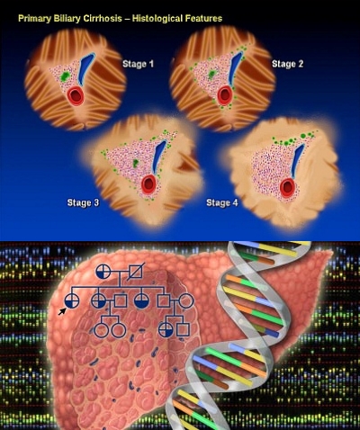
Principal mechanism of liver damage in EPP has been attributed to precipitation of insoluble protoporphyrin in bile canaliculi and to protoporphyrin-induced oxidative stress. The latter arises as a consequence of excess unmetabolized protoporphyrin interacting with the hepatocellular membrane and inducing impaired function of the Na+/K+-ATPase pump within the membrane. The accumulation of excess protoporphyrin, that does not undergo biliary excretion, exacerbates cholestasis and further reduces the excretion of protoporphyrin. These pathophysiological phenomena may result in hepatic inflammation, progressive hepatocellular disease, hepatic fibrosis, and, eventually, cirrhosis.
Pathogenesis and treatment of epatic damage: Liver disease and erythropoietic protoporphyria, 2010
GENETICS AND PHENOTYPES
OMIM
KEGG PATHWAY


In erythropoietic protoporphyria (EPP) there is a mutation of the gene that encodes ferrochelatase (FECH) in the long arm of chromosome 18. This enzyme belongs to the pathway of heme synthesis and catalyzes the insertion of ferrous iron into the protoporphyrin IX ring to form heme ( the last reaction of this pathway ). FECH expression has a heme-dependent negative feedback regulation at a post-transcriptional level in such a way that FECH is decreased by increasing the level of intracellular heme. EPP exhibits both recessive and dominant patterns of inheritance and a high degree of allelic heterogeneity with incomplete penetrance. Most heterozygotes are asymptomatic. Symptoms do not occur unless FECH activity is less than 30% of normal, but such low levels are not present in a majority of patients.
In most patients with EPP, a FECH mutation that markedly decreases or abolishes enzyme activity can be identified on only one allele.
FECH mutations on both alleles are an uncommon cause of EPP. To date, twenty-one symptomatic patients from 17 families have been reported. In the UK and France, this type of EPP accounts for about 4% of EPP families. Reported FECH activities, either measured in lymphocytes or estimated from in vitro expression studies, are within the median range of 9%. Molecular analyses show that most of the patients are compound heterozygotes. In marked contrast to the type of EPP described above, missense mutations make up 85% of the total and null mutations are unusual.
The pattern of inheritance of photosensitivity in these families is characteristic of an autosomal recessive disorder. There is an increased incidence of consanguinity, only siblings are affected and both parents are clinically normal.
Patients in which EPP is inherited in an X-linked dominant pattern have normal FECH activities but higher erythrocyte total protoporphyrin concentrations than other types of EPP of which around 40% is zinc-protoporphyrin. Two frameshift mutations have been identified in 8 families that lead to predicted disruption or deletion of the 19-20 C-terminal amino-acids of ALAS2 (aminolevulinic acid synthase). Prokaryotic expression studies show that both mutations markedly increase ALAS2 activity. Thus the C-terminal region of ALAS2 appears to inhibit enzyme activity; its removal by mutation leads to gain of function whereas all other previously described mutations in ALAS2 decrease activity and cause hereditary sideroblastic anaemia.

Acquired somatic FECH mutations have been identified in a small number of patients in whom EPP has developed after the age of 40 years in association with myelodysplasia or myeloproliferative disorder.
The cutaneous features of EPP, though varying in severity are remarkably uniform. No correlation between indices of severity of photosensitivity, such as age of onset or duration of symptoms, and genotype have yet been reported apart from the suggestion that the relatively common missense mutation, P334L, may cause only mild disease Apart from this instance, no correlation between erythrocyte protoporphyrin concentration and type of FECH mutation has been reported. Seasonal palmar keratoderma has to date been reported only in patients who are compound heterozygotes or homozygotes for FECH mutations.
Focus on genetics: Inheritance in erythropoietic protoporphyria, 2006
Erythropoietic protoporphyria, 2009 Sep
Autosomal recessive disorder, 2010
X-linked Sideroblastic Anemia Due to Carboxyl-terminal ALAS2 Mutations, 2012
CLINICAL DESCRIPTION
FECH deficiency is associated with increased concentrations of protoporphyrin in erythrocytes, plasma, skin and liver.
EPP usually presents in childhood, but there are a few reports of cases presenting in adults.
Skin symptoms: photosensitivity

EPP manifests clinically by skin symptoms of immediate painful photosensitivity. This manifestation starts usually in early infancy or childhood, upon the first sun exposures. Skin stinging, prickling and burning on exposed areas are the initial symptoms experienced by the patient during or shortly after sun exposure. These symptoms may have a variable duration, depending on the intensity and duration of sun exposure. If the exposure has been long enough, erythema and oedema may appear, together with petechiae in some cases. Areas affected are the face and dorsa of the hands, but photosensitivity reactions can also appear on any exposed skin area. Reactions may persist for hours up to several days. The appearance of vesicles or bullous lesions characteristic of other forms of cutaneous porphyria is unusual but may occur. Nail lesions (photoonycholysis and transversal leuconycholysis) are possible associated manifestations.

Late-onset cases have been reported but are exceptional and related to haematologic malignancies, although cases without this association have been described.
Repeated photosensitivity episodes result in altered skin appearance with permanent changes, such as skin thickening with a waxy or leathery appearance, and areas of hyperkeratosis. These lesions are usually located on the dorsa of the hands and face, as these are skin areas usually exposed. The lips show linear furrows and pseudo-rhagades that arte also characteristic. In contrast to other forms of cutaneous porphyria, lesions of milia, hypertrichosis or hyperpigmentation do not appear. This last cutaneous sign may only appear in patients with severe hepatic involvement presenting with cholestasis, itching and jaundice.
Seasonal palmar keratoderma has been reported in some EPP patients and in addition this may indicate autosomal recessive inheritance of the disease.
Hepatobiliary disease
Clinical findings suggestive of liver disease appear in approximately 5%-20% of patients. The susceptibility of individual patients with EPP to protoporphyrin-induced liver damage is highly variable. The host factors responsible for this variable susceptibility are unknown. The spectrum of hepatobiliary disease associated with EPP is wide. It includes cholelithiasis, mild parenchymal liver disease, progressive hepatocellular disease and end-stage liver disease.
Cholelithiasis: Insoluble protoporphyrin in bile may act as nuclei for stone formation. Cholelithiasis is frequent in EPP (10%-20%), due to the accumulation of free protoporphyrin and increased biliary protoporphyrin concentration. Clinical manifestations of cholelithiasis and choledocolithiasis are similar to lithiasis by cholesterol or bilirubin. Many patients with EPP and gallstones undergo cholecystectomy. Subsequent analysis of the stones reveals that they are birefringent and contain high concentrations of protoporphyrin. Some authors believe that EPP should be suspected when cholelithiasis presents in childhood.
Mild parenchymal liver disease: Most patients (20%) present with a mild liver disease, characterized by increased levels of aminotransferases and/or cholestatic enzymes. Typically, in patients with mild disease, there are no symptoms. Patients can also present with splenomegaly and hepatomegaly. Liver biopsy in such patients may reveal features of appreciable hepatocellular injury.
Progressive hepatocellular disease: Symptoms include upper abdominal pain and jaundice. There may be associated rapid deterioration of photosensitivity because of decreased secretion of protoporphyrin into bile, secondary to cholestasis and hemolysis.
It is rare for the initial presentation of EPP to be manifestations of progressive hepatocellular disease. When jaundice is clinically evident, hepatocellular disease is advanced and hepatic clearance function is appreciably reduced. Blood protoporphyrin levels increase further, but fecal protoporphyrin excretion decreases.
End-stage liver disease: Only 5% of liver damage presents as an acute liver insufficiency. Progressive hepatocellular disease ultimately leads to cholestatic hepatocellular failure, which often has an acute onset, and a rapidly progressive, irreversible course. Progressive hepatocellular disease in EPP is usually fatal within months if liver transplantation is not undertaken.
Pathogenesis and treatment of epatic damage: Liver disease and erythropoietic protoporphyria, 2010
Haematological alterations
Microcytic anaemia occurs in 20% to 60% of patients. Erythropoiesis was impaired in most patients with dominant EPP from UK and France [18,19]. All had a downward shift in haemoglobin (Hb), women were slightly more anaemic than men. Iron stores, assessed by serum ferritin (sFn), were decreased by two-thirds, but normal serum soluble transferrin receptor-1 and iron concentrations suggested that erythropoiesis was not limited by iron supply. FECH deficiency in EPP appears to lead to a steady state in which decreased erythropoiesis is matched by reduced iron absorption and supply. This response may in part be mediated by protoporphyrin
Erythropoiesis and iron metabolism in dominant erythropoietic protoporphyria, 2007
DIAGNOSIS
EPP is generally suspected by the presence of acute photosensitivity of the skin and can be confirmed by detection of a plasmatic fluorescence peak at 634 nm. It is also useful finding increased levels of protoporphyrin in feces and the demonstration of an excess of free protoporphyrin in erythrocytes.

Screening for FECH mutation on one allele or aminolevulinic acid synthase 2 gain-of-function mutation in selected family members may be useful, especially in genetic counseling. If one parent is affected with EPP with a FECH allele mutation, the risk of the offspring developing EPP is less than 2.5%, therefore, screening for the presence of the FECH IVS3-48C allele in the other parent may be helpful to estimate the probability for the offspring.
In order to evaluate the hepatic localization of the disease clinicians should explore hepatic function and perform abdominal ultrasonographic to detect cholelithiasis.
Liver biopsy confirms hepatic disease in EPP by the presence of protoporphyrin deposits in the hepatocytes that can be observed as a brown pigment within the biliary canaliculi and the portal macrophages. Macroscopically, the cirrhotic liver can have a black color due to protoporphyrin deposits. The examination of liver tissue under a Wood’s lamp reveals a red fluorescence due to protoporphyrin. Liver biopsy is not helpful for estimation of prognosis of liver disease.
Differential diagnosis
Clinically, EPP should be differentiated from phototoxic drug reactions, hydroa vacciniforme, solar urticaria. Contact dermatitis and angioedema or even other types of curaneous porphyria could also be considered. Chronic lesions should be differentiated from lipoid proteinosis.
Erythropoietic protoporphyria, 2009
MANAGEMENT
Management of photosensitivity
Management of photosensitivity is an essential therapeutic measure and it is based on:
- Avoidance of sun exposure, even through window glass (e.g. car driving). The use of adequate clothing (hats, glasses, gloves) and sunscreens is also advisable. Due to the fact that protection should cover both long-wave radiation plus visible light, physical sunscreens are more effective.
- Topical corticosteroids may be prescribed but are usually ineffective. Oral treatment, if required, may include non-steroidal anti-inflammatory drugs or oral corticosteroids.
- Betacarotene has been used extensively, with the aim of improving light tolerance in EPP patients. Some benefit from its use has been experienced by patients. The following doses of betacarotene are recommended: up to 90-120 mg/day in children and up to 180-300 mg/day in adults. Recent studies, however, have suggested inverse associations between carotenoids and lung cancer mainly in smokers.
- Other treatment approaches include the administration of Vitamin C and E, and cysteine (500 mg/twice daily), orally. Antihistamines may also be used as they may limit the phototoxic reaction.
- UVB/NBUVB phototherapy or PUVA may be used in order to induce tanning and improve the sunlight tolerance. The duration of the treatment is limited by the guidelines of phototherapy and depends also on the patient benefit obtained in reduction of photosensitivity.
- Alfamelanotide (an alpha-melanocyte-stimulating hormone analogue that induces epidermal melanin formation) has recently been shown to have beneficial effects in patients with EPP.
A systematic review of treatment options for dermal photosensitivity in erythropoietic protoporphyria, 2009
Erythropoietic protoporphyria, 2009
α-Melanocyte-stimulating hormone: a protective peptide against chemotherapy-induced hair follicle damage?, 2014
An α-Melanocyte–Stimulating Hormone Analogue in Erythropoietic Protoporphyria, 2009
Management of liver disease and Reduction of protoporphyrin levels
Regarding the reduction of excess protoporhyrn in patients with EPP, two approaches can be considered:
- Reduction of erythropoiesis: this may be achieved by exchange transfusion or hypertransfusion. It is not clear if haematin may temporarily supress erythropoiesis by modifying the activity of the rate-limiting first enzyme of the haem synthetic pathway, ALA synthase. However, this approach cannot be considered as a long-term treatment of the disease.
- Cholestyramine (a bile sequestering agent) may be used to increase the elimination of the excess protoporphyrin through the biliary system. It binds the excess protoprophrin, enhancing its faecal elimination, reducing its plasma and red blood cell concentration, and preventing the protoporphyric hepatopathy. Other similar drugs have been considered, such as chenodeoxycholic acid and ursodeoxycholic acid.
- N-acetyl cysteine: In liver diseases free radicals are increased, thereby damaging the hepatic tissue. In addition, nitric oxide has deleterious effects in the presence of reactive oxygen species, participating in the pathophysiology of different liver diseases.
- Infusions of hematin: Hematin appears to reduce excess protoporphyrin production in the bone marrow. It has been administered to patients with EPP (3-4 mg/kg iv) who develop a crisis after liver transplantation.
- Parenteral iron and transfusion of erythrocytes: the objective of administering iron and/or erythrocytes is to suppress erythropoiesis and, hence, reduce the protoporphyrin level. In theory, iron therapy should not work since it stimulates heme synthesis via 5-aminolevulinate synthase. However, results of this approach in patients with EPP are contradictory. It has been reported that iron therapy may exacerbate hepatic dysfunction whereas in some case reports the correction of iron deficiency has improved EPP. Nevertheless, the mechanism of this favorable response to iron therapy remains unknown and, so, the decision to use this therapy should be individualized.
- Plasmapheresis: Circulating levels of protoporphyrin can be decreased by plasma exchange.
- Extracorporeal albumin dialysis: This type of dialysis is used to decrease circulating levels of albumin-bound protoporphyrin.
- Bone marrow transplantation: The purpose of this approach is to remove the tissue primarily responsible for the overproduction of protoporphyrin. It is a frequently discussed option, but the incidence of associated adverse events has limited its use as a treatment for EPP.
- Liver transplantation: liver transplantation does not correct the constitutional deficiency of FECH. Consequently, there is a risk of recurrence of liver disease after liver transplantation as a result of the continuing overproduction of protoporphyrin.
Liver disease in erythropoietic protoporphyria, 2007
Curative bone marrow transplantation in erythropoietic protoporphyria after reversal of severe cholestasis, 2007
Pathogenesis and treatment of epatic damage: Liver disease and erythropoietic protoporphyria, 2010
Liver transplantation, 2011
Management of microcytic anaemia
Patients studied had a mild microcytic anaemia and thrombocytopenia, as shown by the downward shift of hematologic parameters, which positively correlated with the amount of erythrocyte PPIX. Interestingly, erythropoiesis did not seem to be limited by iron supply in patients, since serum iron and soluble transferrin (Tf) receptor (sTfR) were normal. Therefore oral iron supply is usually found ineffective in trying to correct the usual mild anaemia in EPP patients and is therefore not recommended. However intravenous iron supply or even blood transfusion should be considered in EPP with a more pronounced anaemia.
Erythropoiesis and iron metabolism in dominant erythropoietic protoporphyria, 2007
PROGNOSIS
EPP is a lifelong disease whose prognosis depends on the evolution of the hepatic disease that may lead to potentially fatal liver failure. Regarding life quality aspects, only one study has addressed this question in porphyric patients and, specifically, in EPP patients. It has been concluded that photosensitivity may significantly modify the life style of EPP patients, with a marked impact on the quality of life and with scores that appear higher in EPP patients as compared to those for other skin diseases considered as severe.
Pathogenesis of demage: Erythropoietic protoporphyria, 2009