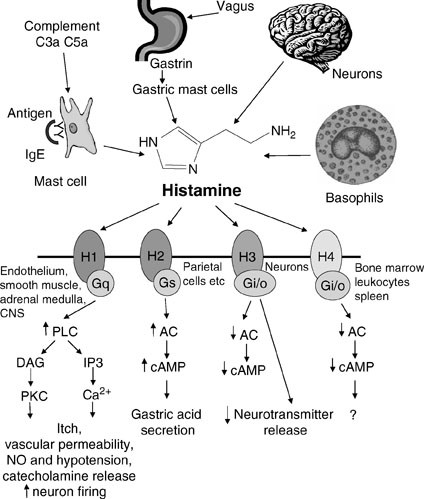
Histamine
Histamine is a signal molecule derived by histidine.
Autacoids: histamine and bradykinin
Histamine metabolism

Histidine Decarboxylase
Bacterial Histamine synthesis
A Potential Central Hub of Histamine in the Microbiota–Gut–Joint Axis in Rheumatoid Arthritis: Mechanisms and Translational Implications, March 2026

Histidine Decarboxylase Expression
Histamine Localization
Histamine-containing peripheral neuronal and endocrine systems. 1985
- Abstract
An immunohistochemical method was developed to detect histamine in tissues. The aim of this study was to reveal the cellular stores of histamine in the gastrointestinal tract, pituitary, and adrenal gland. Histamine-containing nerve fibers were found in both rat and guinea pig gut. The origin of at least some of these fibers in the rat ileum was the submucous ganglion cell layer. In the rat stomach, numerous enterochromaffin-like cells exhibited histamine immunofluorescence, and endocrine cells in the ileum and jejunum contained histamine. Only mast cells contained histamine in the neurohypophysis. A large number of process-bearing cells in the guinea pig but not in the rat adrenal medulla contained histamine. The study shows that histamine is present in peripheral nerves and endocrine cells in addition to mast cells, and may function as a neurotransmitter or hormone.
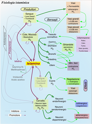
Histamine storage and release
Fluvastatin inhibits mast cell degranulation without changing the cytoplasmic Ca2+ level. 2009
- The statin-induced inhibitory action may be mediated by the suppression of geranylgeranyl transferase via the depletion of intracellular mevalonic acid.
Histamine Targets
Role of Histamine in Modulating the Immune Response and Inflammation, 2018
Histamine Receptors
 ranitidine+and+autism

Role of gastrin in the development of gastric mucosa, ECL cells and A-like cells in newborn and young rats. 2002
- Histamine-producing ECL cells and ghrelin-producing A-like cells are endocrine/paracrine cell populations in the acid-producing part of the rat stomach.
Rat stomach ECL-cell histidine decarboxylase activity is suppressed by ergocalciferol but unaffected by parathyroid hormone and calcitonin. 1999
Motilin Stimulates Gastric Acid Secretion in Coordination with Ghrelin in Suncus murinus. 2015
- BACKGROUND Ghrelin, a growth-hormone-releasing peptide, has two major molecular forms: acylated (acyl) and desacylated (desacyl). Recent studies suggest different roles for these two forms. In the present study, we compared desacyl and acyl ghrelin with regard to acid secretion and histamine production in the rat stomach.
METHODS We performed in vivo experiments using gastric lumen-perfused rats. The effects of the two forms of ghrelin on gastrin (gastrin-17)-stimulated acid secretion were also examined. Furthermore, to examine the effects of ghrelin on histamine production, histidine decarboxylase messenger ribonucleic acid in the gastric corpus mucosa was measured by reverse transcription-polymerase chain reaction.
RESULTS Intravenous administration of acyl ghrelin at 20 μg/kg increased gastric acid secretion to 4.8 times greater than control levels. However, desacyl ghrelin had no effect on acid secretion, even at 200 μg/kg. Acyl ghrelin enhanced gastrin-stimulated acid secretion while desacyl ghrelin did not. Vagotomy markedly inhibited the enhancement of gastrin-stimulated acid secretion by acyl ghrelin. Acyl ghrelin increased histidine decarboxylase messenger ribonucleic acid concentration by 2.3 times compared with basal levels at 1 h after administration and by 2.7 times at 2 h after administration; desacyl ghrelin had no such effect. Synergism between acyl ghrelin and gastrin was seen regarding histidine decarboxylase messenger ribonucleic acid concentration.
Hypothalamic neuronal histamine regulates sympathetic nerve activity and expression of uncoupling protein 1 mRNA in brown adipose tissue in rats. 2004?
- To clarify how hypothalamic neuronal histamine regulates peripheral energy expenditure, we investigated the effect of infusion of histamine into the third cerebral ventricle or discrete hypothalamic regions on sympathetic nerve activity and expression of uncoupling protein 1 (UCP1) mRNA in brown adipose tissue (BAT). Infusion of histamine (200 nmol) into the third cerebral ventricle of anesthetized rats significantly increased the electrophysiological activity of sympathetic nerves (P<0.01) and UCP1 mRNA expression in the BAT (P<0.05). Microinjection of histamine (10 nmol) into the paraventricular nucleus (PVN) and preoptic area (POA) produced similar significant increases in BAT sympathetic nerve activity (P<0.01 for each). By contrast, injection of histamine into the ventromedial hypothalamic nucleus or lateral hypothalamic area had no effect. We conclude that hypothalamic neuronal histamine may regulate energy expenditure in BAT through the activation of sympathetic nerves. The PVN and/or POA appear to be the principal hypothalamic sites that mediate the stimulatory effect of histamine on this efferent pathway.
Targeted disruption of H3 receptors results in changes in brain histamine tone leading to an obese phenotype. 2002
More on histamine..
VEGF, substance P and stress, new aspects: a revisited study. 2010
- Mast cells play an essential role in diverse physiological and pathological processes, such as atherosclerosis, malignancy, asthma, pulmonary fibrosis and arthritis, directly interact with bacteria, and appear to play a vital role in host defense against pathogens. Mast cells could be recruited in the inflammatory site, by MCP-1, RANTES and SCF, to selectively secrete proinflammatory molecules; these could include growth factors, histamine, which is mitogenic (H1) and an immunosuppressant (H2), neovascularization agents, such as heparin, IL-8, and VEGF, as well as proteases that could permit new blood vessel formation. Neurogenic inflammation involves vasodilation and plasma protein extravasation in response to neural stimulation. Upon stimulation, sensory neurons release Substance P and other neuropeptides and activate neurokinin-1 receptors leading to plasma protein extravasation from post-capillary venules. Substance P is a neuropeptide that is released from nerve endings in many tissues and plays an important role in immunological and inflammatory states, and it is also a mediator of tissue injury, asthma, arthritis, allergy and autoimmune diseases. SP-positive nerve fibers and mast cell contacts are increased by acute stress in mice leading to dermal mast cell degranulation. VEGF is produced by flammatory cells. IL-33 is the newest inflammatory member of the IL-1 cytokine family and we show here that SP can induce VEGF secretion from mast cells and IL-33 augments the effect of SP in VEGF transcription and translation protein.
Histamine breakdown
Dynamics of Histamine in the Brain,
Specific enzymes control histamine synthesis and breakdown
Figure 14-3 summarizes the major mechanisms for the synthesis and metabolism of histamine. Biosynthesis is performed in one step by the enzyme l-histidine decarboxylase (HDC, E.C. 4.1.1.22). Histamine metabolism occurs mainly by two pathways. Oxidation is carried out by diamine oxidase (DAO, E.C. 1.4.3.6), leading to imidazole acetic acid (IAA), whereas methylation is effected by histamine N-methyltransferase (HMT, E.C. 2.1.1.8), producing tele-methylhistamine (t-MH). IAA can exist as a riboside or ribotide conjugate. t-MH is further metabolized by MAO-B, producing tele-methylimidazole acetic acid (t-MIAA). Note that histamine is a substrate for DAO but not for MAO. Aldehyde intermediates, formed by the oxidation of both histamine and t-MH, are thought to be quickly oxidized to acids under normal circumstances. In the vertebrate CNS, histamine is almost exclusively methylated and only small amounts of DAO are detectable. However, IAA, a GABA agonist, has been detected and can be formed in the rat brain. Although IAA in the brain is probably normally formed by the transamination of histidine, it can also be formed by histamine oxidation under some circumstances. In some invertebrate nervous systems, such as Aplysia, histamine is metabolized to γ-glutamylhistamine.
Go to:
Several forms of histidine decarboxylase may derive from a single gene
A single cDNA, cloned from rat [16], mouse and human cells, encodes a 74-kDa protein with functional HDC activity. Two species of HDC mRNA have been found in some cells, but the larger one, which contains an additional insert sequence, does not encode a functional enzyme and is not found in brain. Consistent with the immunohistochemical studies discussed above, HDC mRNA is localized to the caudal hypothalamus in rats. The human HDC gene is large, composed of 12 exons with a size of about 2.4 kb. The enzyme, which requires pyridoxal-5′-phosphate as a coenzyme, shares some homology with DOPA decarboxylase (Chap. 12), another enzyme that requires this cofactor. Protein purification studies have shown HDC to be a dimer composed of two identical 55-kDa subunits. Post-translational modification of the enzyme occurs by an elastase-like enzyme, which converts the 74-kDa form to the smaller protein. Biochemical, biophysical and immunological studies have suggested the existence of HDC isoenzymes [2], now thought to result from either post-translational modification of the protein or, possibly, allelic variants. The rat HDC gene has two potential N-glycosylation sites and two recognition sequences for phosphorylation by cAMP-dependent protein kinase (PKA). These potential modulatory sites might contribute to HDC heterogeneity [16].


Histamine - beyond mastcells
Neuronal histamine pathways

Characteristics of Histamine Receptors
THE GENES
Protein Aminoacids Percentage (Width 700 px) of Histamine Receptors


DAO is more ancient than MAO, and depends on very low Oxygen concentration or glucocorticoids (??)


The evolutionary sequence is HDC --> OXDA --> HNMT --> MAOA --> MAOB


Apparently, histamine receptors evolve earlier than HNMT, MAO, pointing to a separate evolutionary pathway

A hypothetical explanation is:
- HR2 senses histamine, a chemical by product of histidine (a danger signal)?
- Evolution leads to enzymatic histamine synthesis (by HDC) and immediately two additional receptors HR1 and HR4 arise.
- Histidine decarboxylase expression
- All further molecules, including HR4, are aimed at reducing the amount of histamine active in the different tissues
SYNTHESIS AND TURNOVER
mRNA synthesis
protein synthesis
post-translational modifications
degradation
Histamine reuptake??
CELLULAR FUNCTIONS
cellular localization,
biological function
- Cell signaling and Ligand transport
- Structural proteins
REGULATION
Cofactor for gaba, dopamine and histamine?

- The pathway of histamine synthesis stimulation and inverse agonist through histidine decarboxylase (HDC). Up-regulation of H1-histamine receptor is induced by IL-4 through the activation of H1-histamine receptor genes and histidine decarboxylase gene. Antihistamines has a mechanism to inhibit the up-regulation of H1-histamine gene expression and suppress histamine basal signal through inverse agonistic activity. Downregulation of H1-histamine gene expression by antihistamines occurs through suppression of histidine decarboxylase and IL-4 gene transcription.
DIAGNOSTIC USE
Somatostatine and histamine
Mainly in gastric cells. What about neurons??