Circadian Rhythms And Metabolism
hai il

Author: Giulia Baldon
Date: 19/01/2013

Description

Origins and functions of the circadian rhythms and their participation in the metabolism.

Biological clocks are genetically encoded oscillators that allow organisms to anticipate changes in the light-dark environment that are tied to the rotation of the Earth.
These “clocks” are expressed throughout the central nervous system and peripheral tissues of multicelled organisms in which they influence sleep, arousal, feeding and metabolism.
In the last decade, researchers start to analyse their function in health and disease at the cellular and molecular level.

Origins of the circadian clocks

The correspondence between biological and geophysical phenomena was not recognized until the early 1700s, when Jean-Jaques d'Ortous De Marain, a french astronomer, demonstrated that the leaves of Mimosa Pudica continue to open and close every 24 hours even when the plant was enclosed in a sealed box.
Since then, nearly all forms of life on the surface of the planet have been shown to exhibit similar circadian cycles, therefore it became necessary defining the characteristics of biological clocks: a persistent and sustained period lenght under constant conditions, entrainment to environmental signals such as light, and stability across wide variations in temperature.
In human, a first understanding of the phenomena was not based on scientific informations but simply on common facts; the so called Chronotypes emerged from the early 20th century, and divided the population in two groups: some people are “larks” and wake up early, whereas others are “night owls” and stay up late, hinting that there is a biological driver of sleep-wake rhythms.
A triumph of modern genetics has been the identification of the molecular pathways that dictate the sleep-wake cycle and other 24-hour-circadian rhythms.

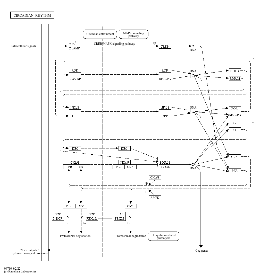
A key advance in circadian genetics was the concept that clocks comprise a transcription autoregolatory feedback loop, with the forward limb encoding activators that promote transcription of a set of repressors, which feed back to inhibit expression and functions.
Indeed, transcriptional oscillators may have provided a selective advantage early in evolution by averting the DNA-damaging effects of sunlight. The presence of a photolyase domain in clock repressors indicates that the timing system co-evolved with DNA repair.

A remarkable series of experiments, such as Light driven changes in energy metabolism directly entrain the cyanobacterial circadian oscillators;2011 , has provided the most convincing evidence for a protein-based clock: a complex of just three protein (KaiA, KaiB, KaiC) together with ATP undergo a self-sustaining 24 hour cycle of alternating phosphorylation and dephosphorylation.
The turnover of ATP modulates the Kai phosphorylation cycle, suggesting that the clock may be coupled to metabolic activity.

An interdependence between circadian and metabolic oscillators has also been suggested by showing that the activity of clock transcription factors is sensitive to redox state; 2011
p=.

More recently, periodic flux in metabolic cycles has been related to production of reactive oxygen species (ROS).

Peroxiredoxin is a redox-sensitive protein, which has a reactive thiol within the active site that is involved in electron transfer from reactive oxygen.
These proteins exhibit 24-hour oscillation in cells, and this cycle is one of the most conserved among species.
Given that eukaryotes first diverged from bacteria around 1.5 billion years ago, a provocative speculation is that oxygenation of the atmosphere conferred ad adaptive advantage on organism with redox-based clock.
In mammals, oscillation in the Prx redox state have been proposed to represent a means of rhytmically anticipating the generation of ROS.
So, if organism are capable of extinguishing ROS, they may have a survival advantage during the oxygen expansion of the atmosphere.
Consideration of the origins of interal clock remains central to our understanding of links between circadian and metabolic systems.

Clocks impact on fitness in multicellular organisms

A principle to emerge from genetic studies is that period lenght is genetically programmed: but how and why are the body's internal clocks set to 24 hours?
Studies such as Plant circadian clocks increase photosynthesis, growth, survival, and competitive advantage;2005 show that bacteria and plants monitored growth and reproduction under conditions in which alignment between “internal clock” and environmental light cycle and show an advantage to period alignment.
So, it became necessary to understand what are the consequences of misalignment at the molecular level: one possibility is that misalignment reduces genome stability by shuffling the phase relationship between cycles of DNA damage and repair, a second hypothesis is that misalignment may superimpose incopatible biochemical processes, such as the oxidative and reductive phases of the metabolic cycle.
For istance, in Saccharomyces cerevisiae , the mutation rate increases when metabolic cycles are misaligned with DNA replication: it is tempting to speculate that the coupling of DNA repair to circadian cycles may contribute to ageing even in higher eukaryotes.

Neural-clock sensory circuit and circadian-system ageing

Understanding how complex organisms detect light and synchronize the clocks in brain and other tissues to environment remains a central challenge in circadian research.
Sensory pathways within the brain have been identified that synchronize the clock independently of visual image formation.
Studies show that even mice without the classic rod and cone visual photoreceptors are still able to synchronize their internal clock to light: the light photoreceptors are expressed in a small number of retinal cells that express the photopigment melanopsin.
Mice that are genetically depleted of these few hundred cells still have normal vision, but they are unable to synchronize their clocks to light: all connections to the hypothalamic pacemakers neurons in the suprachiasmatic nucleus go through these few melanopsin cells.
Suprachiasmatic-nucleus neurons are called the “Master Clock” which synchronizes the hypothalamic control of energy balance, sympathetic outflow and the neuroendocrine systems.

Hypothalamic networks link circadian and energetic centres

The next question is how does the hypothalamic clock communicate with extra-pacemaker and peripheral tissues to produce a coherent phase in the circadian systems throughout the organism?
An appreciation of the central role of the suprachiasmatic nucleus in coordinating sleep-wake behaviour was tested by neural transplantation by using a mutant strain of hamster that shows a short circadian period; 1990 neural grafts from the suprachiasmatic region restored circadian rhythms to arrhythmic animals whose own nucleus had been ablated. The restored rhythms always exhibited the period of the donor genotype regardless of the direction of the transplant or genotype of the host.
These studies also suggested that secreted factors from the suprachiasmatic nucleus contribute to the synchronization of clocks.

Among the areas receiving suprachiasmatic nucleus projections, a large output is toward the lateral hypothalamic area: the nucleus projections synapse on orexin-expressing neurons.
Orexin is neuropeptide that regulates arousal,wakefulness, and appetite: deficiency of either orexin or its receptos is a hallmark of human autoimmune forms of narcolepsy.
Interestingly, narcolepsy is also correlated with elevated body-mass index.
Furthermore, studies such as Low cerebrospinal fluid hypocretin (Orexin) and altered energy homeostasis in human narcolepsy;2001 show that the ablation of orexin receptors increased susceptibilty to died-induced obesity, suggesting that the physiological role of the orexins is to promote arousal and antagonize weight gain.

Ageing and the circadian system

Given the extensive integration of neuroendocrine and circadian systems, it is intriguing to note that suprachiasmatic nucleus function declines with age.
Deficiency of cryptochrome, a repressor of the internal clock repressor, has also been associated with alterations in liver rigeneration, emphasizing the coupling of circadian and cell-cyle pathways.

Cryptochromes are are a class of blue light-sensitive flavoproteins found in plants and animals: the two genes Cry1 and Cry2 code for the two cryptochrome protein CRY1 and CRY2.
In mammals, CRY1 and CRY2 act as light-independent inhibitors of CLOCK - Bmal1 components of the circadian clock.

Circadian origins of metabolic disease

An emerging theme in both circadian and metabolic studies is that it is not only the central nervous system, but also the peripheral tissues that modulate sensory response to the environment.

  • Mounting evidence suggests that alignment between central behavioural rhythms and feeding time is important in metabolic health: e.g. Mice fed with a high-fat diet exclusively during the rest period have accelerated weight gain compared with animals fed with the same diet but during the correct circadian time. Circadian timing of food intake contributes to weight gain;2009

An important goal will be to elucidate the neural and molecular basis of the links between altered timing and behavioural and metabolic disruption: indeed alterations in the time of feeding may induce desynchrony between suprachiasmatic nucleus firing rhythms and imput from peripheral feeding responsive signals.

Function of clock genes in metabolic and vascular disease

Genetic tools to perturb the internal clock have created opportunities to analyse the molecular basis of the clustering of certain pathologies such as morning myocardial infarction and hypertensive crises.
Plasminogen activator inhibitor type 1 or PAI-1 is a major physiologic regulator of the fibrinolytic system and has recently gained recognition as a modulator of inflammation and atherosclerosis. PAI-1 exhibits circadian rhythmicity in its expression, peaking in the early morning, which is associated with increased risk for cardiovascular events, The orphan nuclear receptor Rev-erb alpha regulates circadian expression of plasminogen activator inhibitor type 1; 2006

Both autonomous and non-autonomous vascular effects of the clock cause variation in blood pressure across the ligh-dark cycle; CLOCK expression in the vasculature affects the progression of atherosclerosis, Tissue-intrinsic dysfunction of circadian clock confers transplant arteriosclerosis; 2011

The process of circadian synchronization is analogous to protein folding dynamics, with energy minima across the circadian landscape achieved during phase alignment of individual cells and tissues and misalignment (analogous to misfolding traps) induced by either environmental or behavioural perturbation.

Entraining agents promote synchronization and circadian resonance of individual tissue clocks, whereas circadian insults lead to off-synchrony pathways in which phase and amplitude are misaligned. Such misaligned states may be permanent or re-aligned.

Chronobiology and health and management of disease

The integrative physiology of circadian and metabolic systems has emerged through a combination of biochemical and experimental genetic studies.
Monogenic disorders in sleep onset and waking have provided evidence that clocke genes have an effect not only on subjective chronotype, but also on neurological pathways that regulate sleep in humans.
For example, here there are some implications derived from studies about circadian disorders in humans:

  • A mutation in human Period 2 or hPER2 a gene crucial for resetting the central clock in response to light, is associated with familial advanced sleep phase syndrome (FASPS), an autosomal dominant condition in which patients feel very sleepy and go to bed early in the evening (e.g. 6:00–8:00 p.m.) and wake up very early in the morning (e.g. around 3:00 a.m.). When someone has advanced sleep phase disorder their melatonin levels and core body temperature will cycle hours earlier than the average person.

For extra information about Circadian Sleep Disorders

But maybe the most important results derived from circadian studies are those concerning with metabolic disorders:

For extra information about Cancer and Circadian Rhythms

The Social Jet Lag

The public health implications ,as we can see, may be quite broad given the frequency of circadian behavioural disruption; indeed, the "Social Jet Lag" that is habit of altering bedtime on weekend has been associated with increased body weight, Social Jet Lag and Obesity, 2012

Obesity has reached crisis proportions in industrialized societies, and many factors converge to yield increased body mass index (BMI): among these is sleep duration. The circadian clock controls sleep timing through the process of entrainment. Chronotype describes individual differences in sleep timing, and it is determined by genetic background, age, sex, and environment (e.g., light exposure).

Social jetlag quantifies the discrepancy that often arises between circadian and social clocks, which results in chronic sleep loss.
The circadian clock also regulates energy homeostasis, and its disruption, as with social jetlag, may contribute to weight-related pathologies.

The models included age, sex, and average sleep duration as variables along with social jetlag and chronotype. Analyses confirmed previous findings, showing that age, sex, and sleep duration are important factors in predicting BMI in both weight groups. For individuals in the normal group, social jetlag is not a predictor for BMI (B). In contrast, living against the clock increases BMI in the overweight group, as exemplified by the positive association between BMI and social jetlag. In this case, the influence of social jetlag is more than half of the impact of sleep duration.

These results demonstrate that living “against the clock” may be a factor contributing to the epidemic of obesity.
This is of key importance in pending discussions on the implementation of Daylight Saving Time and on work or school times, which all contribute to the amount of social jetlag accrued by an individual.
Improving the correspondence between biological and social clocks will contribute to the management of obesity.

In conclusion, at this link you can find a simple and clear video about social jet lag.

Comments
2014-01-12T10:42:47 - Alessandra Petitti

Circadian rhythm sleep disorders and role of melatonin

Introduction

Circadian rhythms generated by a central pacemaker, the suprachiasmatic nucleus (SCN) in the anterior hypothalamus, and are synchronized to the external environment .

At the molecular level, circadian clock gene proteins oscillate by means of an autoregulatory feedback loop, generating a self-sustained timing system that is highly regulated with a period of about 24 h . Mammalian Circadian Autoregulatory Loop
An important function of the SCN is to adjust the output signals and the endogenous rhythm in accordance with external time signalling stimuli (ETSS). The afferent connections of the SCN indicate that it is particularly sensitive to light, and light is now considered the most important ETSS. The process by which light synchronizes the SCN to a 24-h day is called entrainment. A human living in a natural habitat will adhere to a 24-h day, primarily due to light exposure.
There exists an important connection between the SCN and the pineal gland. Melatonin, which is the only known hormonal output from the pineal gland, affects the SCN by inhibiting firing. Hence, the SCN and pineal gland seem to be able to influence each other in a mutual way, and play an important role in regulating the sleep-wake cycle.

Sleep regulation

Sleep is regulated by an interplay of different factors main focus has been on the interaction between the homeostatic processes and the endogenous circadian processes.

  • The homeostatic process accumulates as a function of prior wakefulness, there is more homeostatic factor the longer you are awake. This factor is believed to be of main importance for sleep quality; that is, the longer you are awake, the deeper the following sleep episode will be (increased slow wave activity).
  • The circadian factor on the other hand plays an important role in sleep quantity; that is, sleep duration is for the most part determined by when you go to bed. In other words, sleep length is not dependent on the sleep homeostatic factor, but largely dependent on when you go to sleep according to your own circadian rhythm. Night workers have experienced this as their sleep duration is usually shorter (often less than 6 h) than normal when going to bed in the morning, even though they often have been awake for more hours before going to bed than daytime workers.

From a practical point of view, this interaction between the homeostatic and circadian processes means that it is important to be awake for a substantial amount of time to get sleep of high quality, and to have regular bed and rise times in order to have a stable sleep duration.

If there are alterations in the regulation of sleep, insomnia can arise and it is widely studied in Italy by Studio Morfeo: insomnia in primary care, a survey conducted on the Italian population

Core body temperature and endogenous melatonin rhythms

The core body temperature usually peaks in the late afternoon or evening hours, and reaches its lowest point, nadir, in the early morning. Sleep normally occurs on the downward slope of the core body temperature rhythm and normally ends about 2 h following nadir.
A similar coupling between the sleep/wake-rhythm and the rhythmicity of the melatonin secretion is also normally present. Melatonin secretion usually increases soon after the onset of darkness, peaks in the middle of the night and gradually falls during the second half of the night. Sleep usually takes place when the melatonin level is high, and wakefulness normally coexists with low plasma melatonin levels. Based on the close correspondence between sleep/wakefulness and body temperature/melatonin, the core body temperature and melatonin (measured in saliva, urine or plasma) constitute the two most common physiological measures of circadian rhythm.

Melatonin: a major regulator of the circadian rhythm of core temperature in humans,2013

Melatonin : structure, functions and mechanism of action

In humans, the pineal gland lies in the center of the brain, behind the third ventricle.
The gland consists of two types of cells: pinealocytes, which predominate and produce both indolamines (mostly melatonin), peptides (such as arginine vasotocin), and neuroglial cells. The gland is highly vascular.
Melatonin, or N-acetyl-5-methoxytryptamine, was first identified in bovine pineal extracts on the basis of its ability to aggregate melanin granules and thereby lighten the color of frog skin.

Photic information from the retina is transmitted to the pineal gland through the suprachiasmatic nucleus of the hypothalamus and the sympathetic nervous system.

The neural input to the gland is norepinephrine, and the output is melatonin.
The synthesis and release of melatonin are stimulated by darkness and inhibited by light.

  • During daylight hours, the retinal photoreceptor cells are hyperpolarized, which inhibits the release of norepinephrine. The retinohypothalamic–pineal system is quiescent, and little melatonin is secreted.
  • With the onset of darkness, the photoreceptors release norepinephrine, thereby activating the system, and the number of α1- and β1-adrenergic receptors in the gland increases. The activity of arylalkylamine N-acetyltransferase, the enzyme that regulates the rate of melatonin synthesis, is increased, initiating the synthesis and release of melatonin.
    As the synthesis of melatonin increases, the hormone enters the bloodstream through passive diffusion. In humans, melatonin secretion increases soon after the onset of darkness, peaks in the middle of the night (between 2 and 4 a.m.), and gradually falls during the second half of the night. Serum melatonin concentrations vary considerably according to age.

In normal young adults, the average daytime and peak nighttime values are 10 and 60 pg per milliliter (40 and 260 pmol per liter), respectively.

Two membrane-melatonin-binding sites belonging to pharmacologically and kinetically distinct groups have been identified:

  • ML1 (high-affinity [picomolar]) sites; activation of ML1 melatonin receptors, which belong to the family of guanosine triphosphate–binding proteins (G protein–coupled receptors), results in the inhibition of adenylate cyclase activity in target cells. These receptors are probably involved in the regulation of retinal function, circadian rhythms, and reproduction.
  • ML2 (low-affinity [nanomolar]) ML2 receptors are coupled to the stimulation of phosphoinositide hydrolysis, but their distribution has not been determined.

    Melatonin may also act at intracellular sites. Through binding to cytosolic calmodulin, the hormone may directly affect calcium signaling by interacting with target enzymes such as adenylate cyclase and phosphodiesterase, as well as with structural proteins. Melatonin has recently been identified as a ligand for two orphan receptors (α and β) in the family of nuclear retinoid Z receptors. The binding was in the low nanomolar range, suggesting that these receptors may be involved in nuclear signaling by the hormone.

For more information,consult Melatonin in Humans,2007
That substance is critical to adjust the sleep-wake cycle, but plays an important role also in other systemic processes as can be observed from the figure.

Circadian rhythm sleep disorders

An essential role of the circadian clock is to promote wakefulness during the day and, thus, facilitate consolidation of sleep during the night . In humans, the propensity to fall asleep, shows a biphasic circadian rhythm. In most individuals, there is a midday decrease in alertness occurring at around 2–4 p.m., followed by an increase in alertness that peaks during early to mid-evening hours, then it declines to its lowest levels around 4–6 a.m. During the early evening hours, when the homeostatic drive for sleep is high, the circadian alerting signal is also at its highest level .
Circadian rhythm sleep disorders arise from disruption of the circadian timing system or a misalignment between the endogenous circadian timing and the external 24-h social and physical environment, resulting in complaints of insomnia and/or excessive sleepiness and impairment in important areas of functioning and quality of life. The actual clinical presentation of CRSD is often influenced by a combination of physiological, behavioral, and environmental factors.

A clinical approach to circadian rhythm sleep disorders

We haved talked about the dependence of sleep on the biological clock and as it is reflected in the characteristic temporal relationship between sleep propensity and the circadian rhythm of body core temperature.
Under the entrained conditions of normal daily life, this relationship is alteredsomewhat, such that major nocturnal sleep is typically initiated 5-6 h prior to the temperature minimum and is terminated shortly after the minimum. For most people, this corresponds roughly to sleep onset times of between 11p.m. and midnight, and wake-up times of between about 6 a.m. to 8 a.m. Undoubtedly, because the vast majority of humans exhibit similar timing in sleep-wake behavior, societal demands have evolved to accommodate such biological timetables.
For a small percentage of the population, however, there is a misalignment between the endogenous clock that governs the timing of sleep and the sleep-wake cycle that is desired, or which is regarded as the societal norm. These individuals are said to have circadian rhythm sleep disorders .

Delayed sleep phase type (delayed sleep phase disorder, delayed sleep phase syndrome).

Delayed sleep phase disorder (DSPD) is characterized by sleep times that are delayed three to six hours relative to the desired or socially acceptable sleep–wake schedules. Patients typically report difficulty falling asleep before 2–6 a.m. and, when free of social obligations, such as on weekends and vacations, would prefer wake times between 10 a.m. and 1 p.m.

When following socially enforced sleep–wake times, patients will present with classical symptoms of chronic sleep-onset insomnia and difficulty waking up in the morning for work, school or social obligations.

Although the exact pathophysiologic mechanisms of DSPD remain poorly understood, it is likely a result of multiple interacting genetic, physiological and behavioral factors.

  • Suggestion of a genetic basis for DSPD is supported by a report of one large family in which the DSPD phenotype was shown to segregate as an autosomal dominant trait, and recent evidence of polymorphisms in the circadian rhythm genes, such as hPer3, arylalkylamine N-acetyltransferase, HLA and Clock, in individuals with DSPD.
  • Each day the human circadian clock must be synchronized, or entrained to the 24-h geophysical cycle, by being reset earlier (advanced) by 15-20 min. For virtually all organisms, including humans, the natural light-dark cycle provides the strongest such synchronizing signal.
    Individuals with DSPS are, for one reason or another, unable to achieve the usual phase-advance that is required to entrain our longer-than-24-h endogenous clocks to the 24-h day. It may result from a “weak” phase-advance portion of an individual’s PRC to light, or other phase-resetting stimuli.
    It was also observed a prolonged interval from body temperature nadir to sleep offset in patients with delayed sleep phase syndrome
    With respect to the tendency for DSPS to be more frequent in adolescents, Carskadon and co-workers have argued that other changes in the biological timing system- perhaps changes in melatonin output, or gonadotropin secretion-associated with puberty, may be implicated in development of the syndrome.

Advanced sleep phase type (advanced sleep phase disorder, advanced sleep syndrome).

Advance sleep phase disorder is characterized by habitual and involuntary sleep times (6–9 p.m.) and wake times (2–5 a.m.) that are several hours early relative to conventional and desired times . Patients with ASPD typically present with complaints of sleep maintenance insomnia, early morning awakenings and sleepiness in the late afternoon or early evening.
In general, individuals with ASPD tend to have less difficulty adjusting to their preferred earlier schedules than those with DSPD.

Proposed mechanisms include an unusually short endogenous circadian period (less than 24 h) , or changes in the interaction of circadian timing and sleep homeostatic regulation . Furthermore, decreased exposure or weakened responses to entrainment agents such as light and physical activity may also contribute to the advanced sleep phase, particularly in older adults .

Genetic factors are likely to play an important role in ASPD.

Free-running type (non-entrained type, non-24-h sleep–wake syndrome, hypernychthermal syndrome).

Non-24-h sleep–wake syndrome is characterized by a steady daily drift of the major sleep and wake times. Because the endogenous circadian period in humans is usually slightly longer than 24 h, patients will report a progressive delay in the timing of sleep and wake times.

Attempting to maintain a regular sleep–wake schedule can lead to the development of symptoms of insomnia, early morning awakenings and excessive sleepiness that varies in intensity periodically. At times when the endogenous pacemaker is not in phase with the conventional sleep and wake times, patients will report symptoms that cause impairment in social, occupational or other areas of functioning. When the endogenous circadian rhythm is in phase with sleep times, sleep is usually normal.

Irregular sleep–wake type (irregular sleep–wake rhythm)

Irregular sleep–wake rhythm is characterized by the lack of a clearly identifiable circadian pattern of sleep and wake times. Although total sleep time over 24 h may be normal for age, sleep and wake periods occur in short bouts throughout the day and night.

This disorder is most commonly seen in association with dementia, mental retardation and brain injury .

Shift work type (shift work disorder, shift work sleep disorder).

Shift work disorder (SWD) typically presents with complaints of unrefreshing sleep, excessive sleepiness and insomnia that vary depending on the work schedule.

The relationship between sleep problems and work schedule should be evident by the history. SWD is most commonly seen in association with night and early morning (before 6 a.m.) shifts. Patients usually report decreased total sleep time, poor sleep quality and excessive sleepiness at work .
Shift Work Sleep Disorder, Prevalence and Consequences Beyond that of Symptomatic Day Workers

Chronic sleep deprivation may lead to impaired performance at work and safety concerns at work and/or during the commute home SWD results when individuals are required to work and sleep at times that are in opposition to the circadian propensity for sleep and alertness, leading to symptoms of insomnia and excessive sleepiness. Patients may complain of problems initiating and maintaining sleep as they are attempting to sleep at a time of low circadian sleep propensity. Symptoms of insomnia and excessive sleepiness may persist for several days after the last night shift or on days off, even after sleep has been restored to conventional times.

This continued difficulty is likely due to a partial adjustment of the circadian system. Successful adaptation to shift work will be influenced by the number of consecutive night shifts and the speed and direction of the shift rotation.
From the therapeutic point of view were performed different trials on a drug called Modafinil.
For more informations Modafinil for Excessive Sleepiness Associated with Shift-Work Sleep Disorder,2005

Jet lag type (jet lag syndrome).

Jet lag is the result of the external environment being temporarily altered in relation to the timing of the endogenous circadian rhythm by rapid traveling across time zones. It is characterized by symptoms such as daytime fatigue and sleepiness, night-time insomnia, mood changes, difficulty concentrating, general malaise and gastrointestinal problems . During eastward travel, difficulty falling asleep is more prominent, and during westward travel complaint of sleep maintenance is most common.
Symptoms are transient and should resolve as the traveler’s circadian clock re-establishes a normal phase relationship with the local time.

Treatment

The treatment options in ordinary clinical practice for circadian rhythm sleep disorders comprise bright light treatment and exogenous melatonin administration.

Bright light treatment

The SNC is not equally sensitive to the effect of light at all time points during the day, and the type of effect light exposure has on the circadian rhythm is also related to the duration of the light exposure.
Studies, using a variety of experimental designs, have now consistently shown that the effect of light on circadian rhythms follows a so-called phase-response curve (PRC).
A Phase Response Curve to Single Bright Light Pulses in Human Subjects
According to the PRC, light can have two opposite effects on the circadian rhythm.

  • Light exposure before the nadir of the core body temperature rhythm causes a phase delay
  • Light administered after nadir produces a phase advance.
    Thus, light in the evening normally causes phase delay, and light in the morning causes phase advance. Also, light exposure close to the nadir produces the greatest phase shifts. It follows that the further away from nadir light exposure takes place, the less effect it exerts. The magnitude of phase shifts is also a function of the dose and duration of the light exposure. Hence, light can advance or delay the circadian rhythm depending on time of light exposure.
    This therapy is used mainly for ASPD.
    Today, bright light is typically administered by portable units yielding about 10 000 lux, and exposure time is about 30–45 min per day. Studies have also investigated the effects of presenting light of different wavelengths.

The Role of Bright Light Therapy in Managing Insomnia

Visible light with short wavelengths (**blue light**) has a stronger melatonin suppressing effect and a stronger phase shifting effect on the human circadian rhythm compared to light with longer wavelengths.
Light treatment can be self-administered at home, according to a therapeutic regime. Bright light of 10 000 lux for at least 30 min is recommended, but light of less intensity or shorter duration will be better than no light at all.
Furthermore, whenever outdoor light of sufficient intensity is available, being outdoors is preferable to sitting in front of a light box.

It's important to stress as this therapy can be used also to cure some kind of depression as antepartum and postpartum ones. It also used to treat winter depression.
Bright Light Treatment of Winter Depression
Bright Light Therapy’s Effect on Postpartum Depression

Melatonin treatment

Melatonin is rapidly metabolized, chiefly in the liver, by hydroxylation (to 6-hydroxymelatonin) and, after conjugation with sulfuric or glucuronic acid, is excreted in the urine. The urinary excretion of 6-sulfatoxymelatonin (the chief metabolite of melatonin) closely parallels serum melatonin concentrations.
Cytochrome P 450 isoforms involved in melatonin metabolism in human liver microsomes

Intravenously administered melatonin is rapidly distributed (serum half-life, 0.5 to 5.6 minutes) and eliminated. The bioavailability of orally administered melatonin varies widely. Much lower oral doses (1 to 5 mg), which are now widely available in drugstores, result in serum melatonin concentrations that are 10 to 100 times higher than the usual nighttime peak within one hour after ingestion, followed by a decline to base-line values in four to eight hours.
Increasing serum melatonin concentrations (to normal night-time values or pharmacologic values) can trigger the onset of sleep, regardless of the prevailing endogenous circadian rhythm. The hypnotic effect of melatonin may thus be independent of its synchronizing influence on the circadian rhythm and may be mediated by a lowering of the core body temperature.
Melatonin may modify brain levels of monoamine neurotransmitters, thereby initiating a cascade of events culminating in the activation of sleep mechanisms.
In a study of flight-crew members on round-trip overseas flights,56 those who took 5 mg of melatonin orally at bedtime on the day of the return to the point of origin and for the next five days reported fewer symptoms of jet lag and sleep disturbances, as well as lower levels of tiredness during the day, than those taking placebo. However, crew members who started to take melatonin three days before the day of arrival reported a poorer overall recovery from jet lag than the placebo group.
Exogenous melatonin thus appears to have some beneficial effects , mainly for DSPD.
No serious side effects risks have been reported in association with the ingestion of melatonin.

AddThis Social Bookmark Button