written by Chiara Montrucchio and Alessandra Pastorino
DEFINITION
Leishmaniasis (W) is a disease caused by protozoan parasites that belong to the genus Leishmania and is transmitted by the bite of certain species of sand fly, including flies in the genus Lutzomyia in the New World and Phlebotomus in the Old World.

Leishmania (W) are tiny creatures, 3-6 micrometers long by 1.5-3 micrometers in diameter, and found in tropical or temperate regions.
EPIDEMIOLOGY
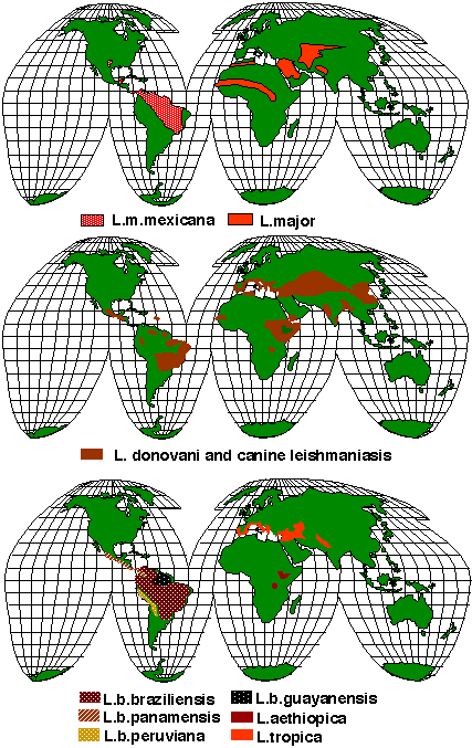
Leishmaniasis can be transmitted in many tropical and sub-tropical countries, and is found in parts of about 88 countries. Approximately 350 million people live in these areas. The settings in which leishmaniasis is found range from rainforests in Central and South America to deserts in West Asia.

More than 90 percent of the world's cases of visceral leishmaniasis are in India, Bangladesh, Nepal, Sudan, and Brazil.
There are a lot of species .
SYMPTOMS
Visceral Leishmaniasis
-the most serious form and potentially fatal if untreated.
Cutaneous Leishmaniasis
-the most common form which causes a sore at the bite site, which heal in a few months to a year, leaving an unpleasant looking scar. This form can progress to any of the other three forms.

| ** | visceral leishmaniasis | Cutaneous leishmaniasis |
|---|
| incubation | weeks to months | weeks to months |
|---|
| symptoms | splenomegaly | papules and nodules |
|---|
| “ | fever | hypo-pigmented macules |
|---|
| “ | hepatomegaly | facial erythema |
|---|
| immunodepression | pneumonia, tubercolosis, dissentery | ** |
|---|
| laboratory | anemia,  plts, plts,  wbc ( wbc (  lymph. ), lymph. ),   IgG, IgG,  albumine albumine | ** |
|---|
Albumin reduction is related to fibrinogen increase, which is used to synthesize globulin by lymphocyte B: A/G ratio is decreased.
So the activation of host immune system is involved in skin ulcer's pathogenesis.
During acute phase response, IL-6 , produced by macrophages, drives liver to produce fibrinogen.
Higher fibrinogen level, both genetic and acquired, determines an increased cardiovascular risk.
*se l'albumina è bassa quale proteina del plasma sarà alta e come questo spiega le ulcere? in realtà è a livello epatico la competizione per gli aminoacidi della dieta e la proteina è il fibrinogeno che è a mio parere usato dai B per fare igG. Ma anche trombosi e tutti i sintomi cutanei. guardate se c'è qualche dato in letteratura su fibrinogeno e il-6. *
Mucocutaneous Leishmaniasis: - commences with skin ulcers which spread causing tissue damage to particularly nose (epistaxis) and mouth
Diffuse cutaneous Leishmaniasis - this form produces widespread skin lesions which resemble leprosy and is particularly difficult to treat.
DIAGNOSIS
Histopathology: amastigotes identification in human tissue (Giemsa)

PCR
laboratory tests: anemia,  plts,
plts,  wbc (
wbc (  lymph. ),
 IgG,
IgG,  albumine
PATHOGENESIS

Leishmaniasis is transmitted by the bite of female phlebotomine sandflies.(1) The sandflie that reach the puncture wound are phagocytized by macrophages (2). Macrophages express a mannose-specific endocytosis receptor that is used by the parasite to penetrate into the host cell.
il mannose-specific endocytosis receptor fisiologicamente a cosa serve? indica che la cellula sta bene, ha tanto ferro o tanto Trp?
Mannose binding lectin pathway can activated complement system , that belongs to the innate immune system. Both high and low iron level
impairs human immune system. Moreover Leishmania required iron for intracellular growth. So, in case of Leishmania's infection, there is a fierce competition between human and parasites to obtain iron.
This mannose receptor synthesis in macrophages is specifically suppressed after infection with Leishmania parasites. Inside the macrophages the parasite transform into amastigotes (3).

Studies indicate that amastigotes of Leishmania use an unusual and unexpected virulence factor, host IgG. This IgG allows amastigotes to exploit the antiinflammatory effects of Fc gamma R ligation to induce the production of IL-10 which renders macrophages refractory to the activating effects of IFN-gamma. Moreover Leishmania can induce PGE2 and COX2 production.
come potrebbe agire la denutrizione? come mancanza di ferro o di aminoacidi essenziali? lamancanza di ferro induce HIF a questo attiva NFKB, forse che lamancanza di Trp impedisce la sintesi di NFKB? c'e' qualcosa su IDO nei macrofagi e Leishmania ?
In condition of malnutrition macrophages, when stimolated with IFN-gamma, produced IL-6, which is a signal of trp's deficiency. In the same condition, NF-kB is also reduced in a first time, and this lead to a reduced NO synthesys.
The same stimolation with IFN-gamma, induced IDO production in macrophages. IDO is the first enzyme responsible of trp cathabolism: tryptophan depletion contributes to the oxygen-independent antimicrobial effects of the activated human macrophage. Infact Leishmania can forbid macrophage's answer to IFN-gamma, hence IDO production.
The endosomes and lysosomes are gradually depleted in iron by host transporters, but pool of intraphagosomal iron is critical for the intracellular survival and replication of these pathogens and so Leishmania amazonensis induces the expression of LIT1 a ZIP family membrane Fe(2+) transporter that is required for intracellular growth and virulence.
Amastigotes multiply in infected cells and affect different tissues: liver, spleen, bone marrow, lymphonodes, skin.(4). These differing tissue specificities cause the differing clinical manifestations of the various forms of leishmaniasis. Sandflies become infected during blood meals on an infected host when they ingest macrophages infected with amastigotes (5,6). In the sandfly's midgut, the parasites differentiate into promastigotes (7), which multiply, differentiate into metacyclic promastigotes and migrate to the proboscis (8)
PATIENT RISK FACTORS
malnutrition
Immunodepression
Immigration and travels have contributed to the spreading of this disease in other countries.
* in realtà una carenza non troppo grave di ferro e una ferritna bassa aumenta i Th1 e potrebbe essere protettivo. Le persone con ferritina alta ammalano di più di lebbra (dati miei da una favela brasiliana) *
TISSUE SPECIFIC RISK FACTORS
physiopathological (due to tissue function and activity): tissues rich in reticuloendothelian cells are specifically involved.
TREATMENT
Treatment usually involves pentamidine or amphotericin B
Amphotericin B associates with ergosterol, a component of fungal membrane, forming a pore that leads to K+ leakage and fungal cell death. The actual mechanism of action may be more complex: the enhancement of macrophage superoxide anion production is supposed.

COMPLICATIONS
HHV8 coinfections
gli HHV8 rubano il ferro come tutti gli herpes ma hanno anche una vIL-6 quindi il problema rimane è più importante la mancanza di ferro o Trp ? vedi
Haemophagocytic syndrome