RAS
Ras-Raf-MEK-ERK-MAPK pathway

Author: yaman hagi
Date: 26/09/2009

Description

DEFINITION

Ras proteins superfamily have served as a prototype for a group of 20-25 kDa guanine nucleotide-binding proteins that share structural homology, the superfamily of Ras-related proteins (Structure of small G proteins and their regulators, 2007). This superfamily of proteins is comprised of over 60 mammalian members and, in terms of both primary sequence and biological activity, it can be conveniently divided into several subfamilies: Ras, Rho, Rab, Arf, Ran and Rad/Gem.

THE GENE

DatabaseLink
HGNCACADM
Uniprot"URL":

CHEMICAL STRUCTURE AND IMAGES

When relevant for the function

  • Primary structure
  • Secondary structure
  • Tertiary structure
  • Quaternary structure


Protein Aminoacids Percentage
The Protein Aminoacids Percentage gives useful information on the local environment and the metabolic status of the cell (starvation, lack of essential AA, hypoxia)

Protein Aminoacids Percentage (Width 700 px)

SYNTHESIS AND TURNOVER

mRNA synthesis
protein synthesis

post-translational modifications
degradation

CELLULAR FUNCTIONS

cellular localization,
biological function

  • Enzymes
DatabaseLink
BRENDA - The Comprehensive Enzyme Information System"URL":
KEGG Pathways"URL":
Human Metabolome Database"URL":
  • Cell signaling and Ligand transport
  • Structural proteins

REGULATION

DIAGNOSTIC USE

DatabaseLink
WikigenesURL
GeneCards"URL":
iHOP"URL":
OMIM"URL":
Entrez Gene"URL":

"WikiGenes":
GeneCards
iHop
OMIM

  • Ras family consists of
    • Ras proteins (H-Ras, K-RasA, K-RasB and N-Ras),
    • four Rap proteins (Rap1A, Rap1B, Rap2A and Rap2B),
    • three R-Ras-like proteins (R-Ras, TC21 and R-Ras3),
    • two Ral proteins (RalA and RalB),
    • the newly identified Rheb and M-Ras proteins.

This family is characterized by high similarity in the effector domain, the so-called switch I region.

CHEMICAL STRUCTURE AND IMAGES

The isoforms are principally distinguished from each other by the final 23–24 amino acid stretch, the hypervariable region (HVR), where there is < 15% sequenze similarity between Ras proteins. The HVR contains all of the motifs responsible for membrane binding and trafficking of each isoform.

Full Size Figure

Protein Aminoacids Percentage

SYNTHESIS AND TURNOVER

After synthesis on cytosolic polysomes (Compartmentalized signalling: Ras proteins and

signalling nanoclusters, 2008
), Ras isoforms undergo a series of posttranslational modifications to increase their membrane affinity.

The cysteine in the C-terminal CAAX motif is farnesylated before the AAX is proteolytically cleaved and the farnesyl–cysteine is carboxymethylated . The farnesyl group promotes weak interaction with the endoplasmic reticulum (ER) which is stabilized by an adjacent set of motifs that vary among Ras isoforms. This consists of mono [N-, K(A)-Ras] or di-palmitoylation (H-Ras) of cysteines or a hexalysine polybasic sequence [K(B)-Ras].The second signal motif and farnesylated cysteineshared by all Ras isoforms comprise the targeting domain; a minimal motif that when fused to GFP displays a superficially equivalent localization as the cognate full length H- and K(B)-Ras proteins. Recent data revealed that a third signal motif is necessary for the correct localization of mono-palmitoylated N- and K(A)-Ras isoforms. GFP conjugated to the minimal targeting domain of N-Ras is restricted to the Golgi, whereas when the adjacent linker region of the HVR is included the construct localizes to the cell surface. When the HVR of the palmitoylated Ras isoforms is compared, there is 70% sequence homology including a six-residue basic ⁄ hydrophobic patch at the N-terminus of the HVR. Mutating this sequence increases the amount of endomembranous localization observed, indicating that this motif contributes to cell-surface localization. Similarly, for K(A)-Ras, the basic patch adjacent to the palmitoyl group is sufficient to ensure cell-surface localization. The second signal motif also determines the trafficking routes taken by H-, N- and K(B)-Ras to the plasma membrane. K(B)-Ras traffics via a poorly characterized Golgi-independent route that in yeast requires class C vps proteins which are normally required to regulate endosome fusion. Unlike other small G proteins such as Rabs and Rho proteins, there has been no chaperone such as GDI characterized for cytosolic Ras trafficking. Although palmitoylated Ras isoforms have also been characterized to traffic via Golgi-independent routes in yeast and adipocytes, in fibroblasts they traffic through the conventional secretory pathway.

The final destination of the post-translationally-modified GTPases depends on the computation
by cells of other ancillary signals present in the GTPase C terminus. In the case of
palmitoylated GTPases, one of these additional signals is the nature of the isoprenyl group
attached to the CAAX box.
Mapping the nucleotide and isoform dependent structural and dynamical features of Ras proteins, 2009 The Ras superfamily at a glance, 2005
Ras Isoform-Specific Signaling: Location,2001)

Like other G proteins, Ras cycles between the GDP-bound inactive form and the GTP-bound active form(Blocking Oncogenic Ras Signaling for Cancer Therapy, 2001).


CELLULAR FUNCTIONS

Ras GTPases—H-Ras, N-Ras, and K-Ras 4B/4A—operate as key molecular switches that convey extracellular signals (Blocking Oncogenic Ras Signaling for Cancer Therapy, 2001) from surface receptors to the interior of the cell, thereby regulating essential processes including proliferation, differentiation, and survival . It is well known that Ras must be attached to the inner leaflet of the plasma membrane protein (PM) ( Na+/K+-ATPase-mediated signal transduction and Na+/K+-ATPase regulation, 2008) to be functional.

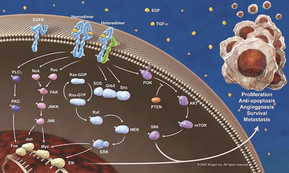
In many cell types, the MAPKs ERK1/2 are linked to cell proliferation (The roles of MAPKs in disease, 2008). ERK1/2 are thought to play a role in some cancers, because mutations in Ras and B-Raf, which can activate the ERK1/2 cascade.In human cancer ( Blocking of p53-Snail Binding, Promoted by Oncogenic K-Ras, Recovers p53 Expression and Function, 2009), the oncogenic mutation of Ras family genes including H-, N-, and K-Ras, is frequently detected in particular, K-Ras mutation is frequent event in

The major alteration known interests the methylation (DNA methylation-mediated nucleosome dynamics and oncogenic Ras signaling, 2008)and on other levels of ras pathway that involve in some somatic gene mutations (Combinatorial patterns of somatic gene mutations in cancer ,2008).

REGULATION

Ras biological activity is controlled by a regulated GDP/GTP cycle. The intrinsic GDP/GTP exchange and GTP hydrolytic activity of Ras is very low and hence, cellular control of GDP/ GTP cycling modulated by two types of regulatory proteins. Guanine nucleotide exchange factors (GEFs; RasGRF/mCDC25, SOS1/2) promote formation of the active GTP-bound state and Ras GTPase activating proteins (GAPs; p120 GAP, NF1-GAP/neurofibromin) promote formation of the inactive GDP-bound state (Increasing complexity of Ras signaling, 1998)

DIAGNOSTIC USE

Naturally as you immagine the ras pathway rappresents a “molecular target”. This is important expecially in the cancer therapies infact the decision to use some drugs are based on the activation or not of the single pathway. (A gene expression predictor of response to EGFR-targeted therapy stratifies progression-free survival to cetuximab in KRAS wild-type metastatic colorectal cancer, 2009,, Novel Agents in CML Therapy: Tyrosine Kinase Inhibitors and Beyond, 2008)

Comments
2021-04-08T21:45:06 - Gianpiero Pescarmona

GTPase cycling between GTP-bound "ON" state and GDP-bound "OFF" state. Upon an external signal, a guanine nucleotide exchange factor (GEF) is activating small GTPases (here H-RAS) by exchanging GDP to GTP and thereby modulating cell signaling cascades. To return into the "OFF" state, GTPase-activating proteins (GAPs) accelerate GTP hydrolysis to GDP.

RAS signaling

  • RAS has no Trp
    • relationships with TNF and IL6 (role in fibrinogen production?)
      • TNF alpha-->HRas-->IL6 and PGH2
      • HRas -- STAT3 phosphorilation--> fibrinogen (Trp supply)?
  • Membrane translocation: farnesylation / myristoylation?

AB0134 Affibody Molecules Inhibiting the Interaction between RAS and RAF Suppress the Proliferation and the Production of Inflammatory Mediators by Synovial Cells 2014

We also demonstrated that signaling through the H-Ras–Raf pathway by MH7A cells can be blocked, thereby inhibiting the productions of IL-6 and PGE2 stimulated by TNF-alpha.

HRAS+and+il6

HRAS+and+stat3

15-Keto prostaglandin E2 suppresses STAT3 signaling and inhibits breast cancer cell growth and progression. 2019

In this study, we observed that 15-keto PGE2 suppressed the phosphorylation, dimerization and nuclear translocation of signal transducer and activator of transcription 3 (STAT3) in human mammary epithelial cells transfected with H-ras (MCF10A-ras).

HRAS+and+aging

H Ras induces cellular senescence. How?

Biological consequences of cellular senescence. Cellular senescence can be induced by various triggers, including, but not limited to, telomere dysfunction, oncogene activation, reactive oxygen species and cell–cell fusion. Short-term presence of senescent cells plays a beneficial role in tumor suppression, wound healing and embryonic development. However, the long-term persistence of senescent cells in tissues can paradoxically promote tumorigenesis and the development of age-related diseases

Physiological and pathological consequences of cellular senescence, 2014

Ras signaling in aging and metabolic regulation, 2017

Aberrant signal transduction downstream of the Ras GTPase has a well-established role in tumorigenesis. Mutations that result in hyperactivation of Ras are responsible for a third of all human cancers. Hence, small molecule inhibitors of the Ras signal transduction cascade have been under intense focus as potential cancer treatments. In both invertebrate and mammalian models, emerging evidence has also implicated components of the Ras signaling pathway in aging and metabolic regulation. Here, I review the current evidence for Ras signaling in these newly discovered roles highlighting the interactions between the Ras pathway and other longevity assurance mechanisms. Defining the role of Ras signaling in maintaining age-related health may have important implications for the development of interventions that could not only increase lifespan but also delay the onset and/or progression of age-related functional decline.

Ras signaling from plasma membrane and endomembrane microdomains, 2005

The localization of Ras isoforms to distinct plasma membrane microdomains is determined by both protein/lipid and protein/protein interactions. Both palmitoylated Ras isoforms undergo differential lateral segregation into discrete microdomains in response to GTP-loading. In the GDP conformation, H-Ras is resident in cholesterol-dependent clusters, but upon GTP-loading, it is recruited to cholesterol-independent domains where it is able to interact with the lectin galectin-1, which presumably stabilizes these domains.

Aberrant signal transduction downstream of the Ras GTPase has a well-established role in tumorigenesis. Mutations that result in hyperactivation of Ras are responsible for a third of all human cancers. Hence, small molecule inhibitors of the Ras signal transduction cascade have been under intense focus as potential cancer treatments. In both invertebrate and mammalian models, emerging evidence has also implicated components of the Ras signaling pathway in aging and metabolic regulation. Here, I review the current evidence for Ras signaling in these newly discovered roles highlighting the interactions between the Ras pathway and other longevity assurance mechanisms. Defining the role of Ras signaling in maintaining age-related health may have important implications for the development of interventions that could not only increase lifespan but also delay the onset and/or progression of age-related functional decline.

The signal transduction from Ras isoforms is compartmentalized both at the cell surface and through differential interaction with intracellular membranes. The dynamic nature of the palmitate modification allows signal transduction from N-Ras and H-Ras to be delicately tuned presumably in response to the nature of the stimulus, allowing signal transduction from the plasma membrane and intracellular compartments. Activation at the plasma membrane results in the activation the Raf/MEK/ERK pathway through recruitment of scaffolding proteins such as KSR in response to the generation of a favorable microenvironment resulting in the rapid generation of a transient signal. H-Ras is also known to be associated with endosomes possibly through the passive internalization with growth factor receptor complexes or through the active recruitment to these vesicles. Interaction of H-Ras with Raf and the endosome-specific scaffolding protein p14/MP1 on this compartment may result in the generation of unique signal output. Activation at the plasma membrane with subsequent depalmitoylation results in the translocation of N-Ras and H-Ras from the plasma membrane to the Golgi complex. Raf is recruited to the Golgi complex and a delayed yet sustained signal can be generated from this compartment presumably through the interaction with Golgi-specific scaffolding proteins such as Sef.

FKBP12 Binds to Acylated H-Ras and Promotes Depalmitoylation, 2011

Dynamic Palmitoylation Links Cytosol-Membrane Shuttling of Acyl-protein Thioesterase-1 and Acyl-protein Thioesterase-2 with That of Proto-oncogene H-Ras Product and Growth-associated Protein-43, 2013

RAS activate purine biosynthesis and not DNA synthesis? more RNA?

RAS+and+purine+biosynthesis

RAS+and+mtor

RAS activates mTOR: RNA and proteins synthesis

PKC is the real activator of DNA synthesis

RAS+and+DNA+synthesis

VEGF activates protein kinase C-dependent, but Ras-independent Raf-MEK-MAP kinase pathway for DNA synthesis in primary endothelial cells. 1999

These results suggest that in primary endothelial cells, VEGF-induced activation of Raf-MEK-MAP kinase and DNA synthesis are mainly mediated by PKC-dependent pathway, much more than by Ras-dependent or PI3 kinase-dependent pathway.

AddThis Social Bookmark Button