DEFINITION
Takayasu’s arteritis is granulomatous vasculitis of the aorta and its main branches which predominantly affects Asian patients younger than 40 years of age. There are different
types of this pathology depending on the aortic tracteffected. The first case of this arteritis has been described in 1908 by Takayasu Mikito doctor.
Generally, vasculitis are characterized by an inflammatory process affecting the wall of
blood vessels which cause not only a damage of the integrity of the vessel, but also an alterations of the blood flow. It is possible to classify the different species of vasculitis according to the type and the diameter of vessels.
EPIDEMIOLOGY
Takayasu's arteritis is much rarer than other forms of arteritis withan incidence of 2.6
patients per million persons / year.Its eziogenesis still remains uncertain. Infectious agents,autoimmune diseases and genetic predisposition may play an important role in the development of this inflammatory disease of large vessels.
PATHOGENESIS
Concerning the histological point of view, the AT has an acute phase and a chronic phase. In the first phase it occurs a panarteritis with inflammation of the “vasa vasorum” of adventitia, followed by a lymphocytic infiltration of the media and finally, an invasion of smooth muscle cells and fibroblasts which thicken the intima. In the chronic phase adventitial fibrosis prevails ,with the destruction of the elastic cells of media and the thicken of the intima. The latency period between the first and the second phase can vary from several months to several years.
Lymphocytes are the main inflammatory cells: the second
some authors would have involved a mechanism of immune cell types predominantly
Th2, capable of activating the production and the recruitment of several interleukins.
These cytokines modulate, in turn, the immune response and activate the processes
of cell death, resulting in apoptosis of smooth muscle cells and playing a
also in the increased role of MMPs and inhibitors of MMPs.
Even macrophages represent important inflammatory cells of the aorta, since they have the
ability to secrete collagenase and elastase, and cytokines play a central role in recruiting
other inflammatory cells, in stimulating the production of other cytokines and in "induce the
lymphocyte differentiation and neovascularization.
A thoracic T cells are the predominant inflammatory cells, followed by
macrophages: in this location would be predominantly a Th1 type immune response, acting through
activation of the IFN-γ and chemokines related to it.
SYMPTOMS
The clinical picture is characterized by:
1. systemic symptoms:they dominate the "pre-pulseless" and may precede months or even years the diagnosis of TA:
fever;
general malaise;
weight loss;
symmetric polyarthritis.
2.vascular symptoms: these symptoms dominate the so called the late stage or occlusive stage ("pulseless") of the TA, and usually vary, depending on the affected districts:
claudicatio intermittens of upper or lower limbs;
masticatory claudication;
ischemic cerebral lesions;
headache;
angina pectoris;
myocardial infarction;
abdominal pain;
dizziness.
However it prevails a symptomatology due to the development of stenosis, occlusion or aneurysm formation in the aortic arch and load its main branches, of 'the thoracic aorta and / or abdominal aorta and arteries lung and is manifested by symptoms of organ damage secondary to the territory involved stricture or aneurysm.
The most characteristic finding goal is the reduction or absence of arterial pulses can also detect peripheral murmurs. It is also possible to find whispers at the level of one or of both carotid arteries or subclavian, and difference of the systolic blood pressure between both arms of 10 mmHg.
Improved Prognosis of Takayasu Arteritis Over the Past Decade, 02-02- 2012
DIAGNOSIS
Laboratory tests show high levels of nonspecific inflammation high level of:
VES;
PCR ;
alpha 1 and alpha 2 globulin;
fibrinogen,leukocytosis with neutrophilia;
platelets;
the hypergammaglobulinemia is common.
Less common are: reduction of complement, positivity of rheumatoid factor and cryoglobulins presence.
Some research has been based on the measurement of circulating levels of matrix metalloproteinases 2 (MMP2; 120 360), 3 (MMP3; 185 250)and 9 (MMP9; 120 361) in patients with Takayasu’s arterite .Metalloproteinases are a family of enzymes whose function is the main cause of extracellular matrix proteins’s degradation, such as collagen, laminin, fibronectin, elastin and the protein section of proteoglycans; they also occur in many different malignancies.Levels of all 3 metalloproteinases were higher in
patients with active disease than in controls and MMP2 levels remained elevated even in remission. In contrast, an improvement in clinical signs and symptoms was associated with a marked reduction in circulating MMP3 and MMP9 levels in all patients.The researchers concluded that MMP2 could be helpful in diagnosingTakayasu arteritis and that MMP3 and MMP9 could be used as activity markers for the disease.
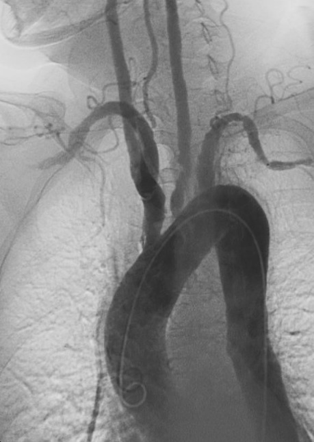
Among the instrumental tests are particularly useful is angiography which can show aneurysmal dilatation. Sometimes both at early stages and late one evidence of the aneurysm may be missing, or because it is still being formed, or because, as may occur in the forms of long duration of treatment, it regressed spontaneously in the fibrous form. The angiography is the "gold standard" for evaluating vascular lesions, in the particular the panangiography allows a correct estimation of the extent of disease, which correlates directly with its severity.
The subclavian artery is the most commonly affected vessel (the left over right), followed in order from the aorta, the common carotid, the renal arteries and the vertebral one.
Less common is the involvement of coronary, pulmonary, iliac, mesenteric.
Important is the ascertainment of renal biopsy even if it not always demonstrate the characteristic lesions of vasculitic.
The histological examination of the vessel wall has a limited role as the biopsy material is available only at the intervention of revascularization, usually performed in the advanced stages of disease.
Ecography also allows to obtain information about the structure of the arterial wall. In recent times also PET seems to have been used not only for early detection but also for assess disease activity.
THERAPY
Mortality at five and ten years is low, with survival rates between 70% and il100%. Among the most frequent causes of death there are congestive heart failure and celebrovascular complications. The goal of treatment is to stop the inflammation arteries and generally this is possible by using high doses of immunosuppressants drugs. Almost half of people with this arteritis draws benefits with corticosteroids (eg prednisone).
After a good initial response, the disease may, however, rekindled and many patients must take cortisone together with traditional immunosuppressants ,as adjuncts to a steroid therapy.
In cases of resistance to traditional therapy it is possible to use biologic. The most analysed one are TNF blockers, although a molecule that blocks IL-6 seems to work better. Interleukin 6 is a protein molecule produced by various kinds of cells, particularly those such as immunocompetent T LYMPHOCYTES and MACROPHAGES in response to trauma or tissue damage. During quiescence periods they can be carried out interventions for the permanent correction of vascular abnormalities in patients at risk for organ injury.
Revascularization may be early, surgical or conduct with percutaneous intraluminal angioplasty techniques. A particular indication for treatment is represented by the presence renovascular hypertension. In recent years Adequate immunosuppression and the selective correction of vascular damage have been much improved the prognosis of Takayasu's disease. Currently good results can be obtain by taking a drug already used for the prevention of rejection in organ transplantation, the micofelonato mofetil.