Ubiquitination
Proteasome

Author: Gianpiero Pescarmona
Date: 17/03/2009

Description

Ubiquitin

Ubiquitin : Exists either covalently attached to another protein, or free (unanchored).

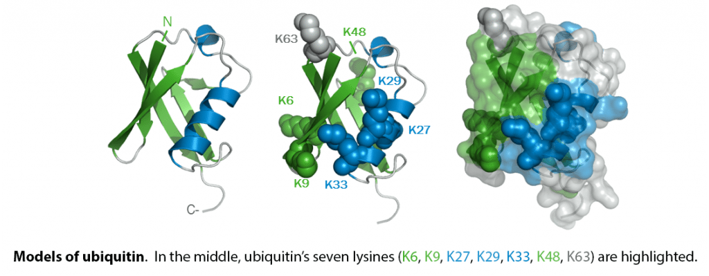
When covalently bound, it is conjugated to target proteins via an isopeptide bond either as a monomer (monoubiquitin), a polymer linked via different Lys residues of the ubiquitin (polyubiquitin chains) or a linear polymer linked via the initiator Met of the ubiquitin (linear polyubiquitin chains). Polyubiquitin chains, when attached to a target protein, have different functions depending on the Lys residue of the ubiquitin that is linked: Lys-6-linked may be involved in DNA repair; Lys-11-linked is involved in ERAD (endoplasmic reticulum-associated degradation) and in cell-cycle regulation; Lys-29-linked is involved in lysosomal degradation; Lys-33-linked is involved in kinase modification; Lys-48-linked is involved in protein degradation via the proteasome; Lys-63-linked is involved in endocytosis, DNA-damage responses as well as in signaling processes leading to activation of the transcription factor NF-kappa-B. Linear polymer chains formed via attachment by the initiator Met lead to cell signaling. Ubiquitin is usually conjugated to Lys residues of target proteins, however, in rare cases, conjugation to Cys or Ser residues has been observed. When polyubiquitin is free (unanchored-polyubiquitin), it also has distinct roles, such as in activation of protein kinases, and in signaling.

from

Ubiquitin is encoded by 4 different genes. UBA52 and RPS27A genes code for a single copy of ubiquitin fused to the ribosomal proteins L40 and S27a, respectively. UBB and UBC genes code for a polyubiquitin precursor with exact head to tail repeats, the number of repeats differ between species and strains.

List of target proteins

  • VHL and p53

BRCA1 Control of Steroid Receptor Ubiquitination 2007

BARD1

A protein that interacts with the N-terminal region of BRCA1 (BRCA1-associated RING domain-1)

Viral activation of ubiquitination

Cellular protein degradation pathways can be utilized by viruses to establish an environment that favors their propagation. Here we report that the Kaposi's sarcoma–associated herpesvirus (KSHV)-encoded latency-associated nuclear antigen (LANA) directly functions as a component of the EC5S ubiquitin complex targeting the tumor suppressors von Hippel-Lindau (VHL) and p53 for degradation. We have characterized a suppressor of cytokine signaling box-like motif within LANA composed of an Elongin B and C box and a Cullin box, which is spatially located at its amino and carboxyl termini. This motif is necessary for LANA interaction with the Cul5–Elongin BC complex, to promote polyubiquitylation of cellular substrates VHL and p53 in vitro via its amino- and carboxyl-terminal binding domain, respectively. In transfected cells as well as KSHV-infected B lymphoma cells, LANA expression stimulates degradation of VHL and p53. Additionally, specific RNA interference–mediated LANA knockdown stabilized VHL and p53 in primary effusion lymphoma cells. Thus, manipulation of tumor suppressors by LANA potentially provides a favorable environment for progression of KSHV-infected tumor cells.

LANA is predicted to form a complex with Cul5/Rbx1 that interacts with Elongin BC but not LANA ΔSOCS (Cul box and BC box) mutant. LANA acts as adapter to link substrates which bind at its amino (1) or carboxyl (2) -terminal domain ( like VHL and p53 ) to EC5S ubiquitin complex and induces the pathway of ubiquitin E1 activation, E2 conjugation, and substrate polyubiquitylation as well as 26S proteasome–mediated degradation.

EC5S Ubiquitin Complex Is Recruited by KSHV Latent Antigen LANA for Degradation of the VHL and p53 Tumor Suppressors 2006 Fulltext

Comments
2010-01-26T10:31:45 - Paolo Fornaciari

Paolo Fornaciari e Giulia Cossu

DEFINITION

The ubiquitin system of protein modification is a crucial mechanism involved in the regulation of a wide array of cellular processes and in the development of various human diseases.
The process of marking a protein with ubiquitin (ubiquitylation or ubiquitination) consists of a series of steps which involve:

  • E1 ubiquitin-activating enzyme
  • E2 ubiquitin-conjugating enzyme
  • E3 ubiquitin ligase.

In the ubiquitination cascade E1 can bind with dozens of E2s which can bind with hundreds of E3s in a hierarchical way.
The resulting modification of the proteins sends them to the proteasome for the degradation.

databaseLink
Kegg URL
WikigenesURL
UniprotURL
Ub (good cross-databases links)E1E2E3
The ubiquitin proteolityc system 2008 YuoTubeURL

CHEMICAL STRUCTURE AND IMAGES

Ubiquitin

Steps

Protein modifiers which can be covalently attached to target lysines either as a monomer or as a lysine-linked polymer. Attachment to proteins as a Lys-48-linked polymer usually leads to their degradation by proteasome . Attachment to proteins as a monomer or as an alternatively linked polymer does not lead to proteasomal degradation and may be required for numerous functions, including maintenance of chromatin structure, regulation of gene expression, stress response, ribosome biogenesis and DNA repair (see below).
The ubiquitin system is a highly regulated enzymatic cascade in which a ubiquitin-activating enzyme (E1) activates the 76 amino acid protein UBIQ (ubiquitin) in an ATP-dependent manner and transfers it to the active site cysteine of ubiquitin-conjugating enzymes (E2s) . Ubiquitin ligases (E3s) have a central role in the process of protein modification with UBIQ (known as 'ubiquitination' or 'ubiquitylation'): they recognize specific substrates and facilitate UBIQ transfer from the E2 onto the substrate. Although the precise number of human E3s is unknown, about 500 or more have been proposed to exist , supportive of the broad role for the ubiquitin system in regulating diverse cellular processes.
We want to underline that E1 needs ATP-Mg2+ for its reaction (till what extents could Mg regulate the activity of this enzyme? Pleiotropic effects of ATP.Mg2+ binding in the catalytic cycle of ubiquitin-activating enzyme.).

Ubiquitin-like proteins (UBLs) have also been identified with varying degrees of identity to UBIQ and are conjugated onto proteins through similar enzymatic cascades as UBIQ (e.g. NEDD8 ) (could be useful another file on this).
Numerous deubiquitylating enzymes (DUBs) have roles in processing polyubiquitin precursor proteins and may also have regulatory roles, e.g. counteracting the ubiquitylation of a particular protein by its cognate E3 and/or proofreading synthesized UBIQ chains. There are also emerging roles for DUBs in disease (could be useful another file on these).
Two major classes of E3s have been identified:
HECT domain E3s form a catalytic UBIQ intermediate on a conserved cysteine residue prior to covalent UBIQ transfer and
the second class of E3s, which contains RING-type and structurally related ligases, facilitates the direct transfer of UBIQ from E2 onto substrate. In general, E3s facilitate covalent UBIQ transfer by properly positioning the site to be modified.
Lysine residues appear to be major sites of UBIQ attachment on proteins, although N-terminal and cysteine modifications have also been reported .

KEGGhsa04120

Feature of the E1 reaction:

Throughout this mechanism, the E1 enzyme is bound to two ubiquitin molecules. Although this secondary ubiquitin is similarly adenylated, it does not form the same thioester complex described previously. The function of the secondary ubiquitin remains largely unknown, however it is believed to facilitate conformational changes observed in the E1 enzyme during the transthioesterification process.

Forms of ubiquitination

A) The ubiquitin modification has three general layouts: mono-ubiquitination, multi-mono-ubiquitination and polyubiquitination. B) Forms of homotypic polyubiquitination, where each ubiquitin chain contains a single linkage type. Individual linkages may lead to distinct ubiquitin chain structure, and Lys48- and Lys63-linked/linear chains have different conformations. We do not know structures of the remaining chain types. Multiple homotypic ubiquitin chains on the same substrate are possible. C) Forms of heterotypic polyubiquitination. In mixed linkages, a ubiquitin chain has alternating linkage types. In branched or forked polyubiquitin chains, a single ubiquitin is extended at two or more lysine residues. (Biochem Soc Trans. 2009 Oct;37(Pt 5):937-53. The emerging complexity of protein ubiquitination. Komander D)

A VERY USEFULL TOOL TO PREDICT UBIQUITINATION SITE : CLICK HERE

IS NECESSARY PREPARE THE SEQUENCE IN FASTA (SOMETIMES READY ON DBs LIKE UNIPROT. OTHERWISE USE WORKBENCH!)

KEY CONCEPT: UPS can recognize or not a sequence of the primary structure of a protein in different moment of the target’s life according to biochemical modifications (i.e. when the proteins are oxidized, or according to particular phase of transduction or post-transductional processing (as phosphorilation). The same way the UPS can ubiquitinate protein before their complete formation (still linked to the ribosome!) or can manage the concentrations of several proteins that need rapid changes of their concentration to fit to the new environment or to mediate a biological response.(could this system regulate some Oscillations? open question)

Proteins aminoacid percentages
The ubiquitin protein itself consists of 76 amino acids and has a molecular mass of about 8.5 kDa. Key features include its C-terminal tail and the 7 Lys residues. It is highly conserved among eukaryotic species: Human and yeast ubiquitin share 96% sequence identity.
E1 is around 60aa, E2 around 150aa and E3 is quite variable but usually 800aa.
We have chosen three representatives of this 3 categories (see above “database: uniprot” for further informations).

Curiously L (highly represented here) regulates the transcription of E3 enzyme (see Muscle Nerve. 2010 Jan 15. Leucine attenuates skeletal muscle wasting via inhibition of ubiquitin ligases. Baptista IL, Leal ML, Artioli GG, Aoki MS, Fiamoncini J, Turri AO, Curi R, Miyabara EH, Moriscot AS.)
(Aminoacids)
As in the proteasomes subunits, W is quite low.
Great gap from Ub to E3 in Q level (activation in different conditions? Check Km and cofactor would be really interesting (open question).

However, ubiquitin is believed to have descended from prokaryotic proteins similar to ThiS or MoaD: these prokaryotic proteins, despite having little sequence identity (ThiS has 14% identity to ubiquitin), share the same protein fold and also share sulfur chemistry with ubiquitin. MoaD, which is involved in molybdenum cofactor biosynthesis, interacts with MoeB, which acts like an E1 ubiquitin-activating enzyme for MoaD, strenghtening the link between these prokaryotic proteins and the ubiquitin system. In the animals (in plants also other roles) Mo is necessary for the degradation of purine and the yelding of uric acid. In some animals the integration of the diet with Mo improve the growth (could UPS regulates this pathway? open question).

SYNTHESIS AND TURNOVER

Ub: Subcellular location, cytoplasm and nucleus (like protesomes).
Post-translational modification: several types of polymeric chains can be formed, depending on the lysine used for the assembly (see above). Ubiquitin is synthesized as a polyubiquitin precursor with exact head to tail repeats, the number of repeats differs between species and strains. In some species there is a final amino-acid after the last repeat while in humans we can observe a Val (meaning of this? Open question). Some ubiquitin genes contain a single copy of ubiquitin fused to a ribosomal protein (either L40 or S27a). Ubiquitin is recycled after the target is commited to the proteasome.

The E1 gene informs us that the Y chromosome derived from the X chromosome!

(see this interesting work:
Hum Mol Genet. 1998 Mar;7(3):429-34.The origin and loss of the ubiquitin activating enzyme gene on the mammalian Y chromosome )

CELLULAR FUNCTIONS:

1.The stability of P53 (p53) is regulated by ubiquitin ligases and a deubiquitylating enzyme
2.Cullin-RING ubiquitin ligases and the APC/C regulate cellular proliferation
The protein encoded by the tumor suppressor gene VHL (von Hippel-Lindau) serves as a substrate receptor for a CUL2-based ubiquitin ligase . Mutations in VHL are associated with lung cancer, sporadic clear cell renal carcinomas and an autosomal dominant familial cancer known as von Hippel-Lindau disease . Many of these mutations prevent VHL associating with the other subunits of its ubiquitin ligase. A substrate for this ubiquitin ligase is a marker of tumor hypoxia, the transcription factor HIF1A (HIF1α, hypoxia-inducible factor 1α ), which stimulates angiogenesis . Numerous biochemical and structural studies have determined that HIF1A binds to VHL when hydroxylated on two proline residues through the activity of prolyl hydroxylases, which results in its ubiquitylation and ultimate degradation .
(renal cancer , looking at this pathway, should one think what are natural regulators of the UPS? Could we use that to change the natural course of these disease? open question). Linked to the HIF argument there is also the wound healing
3.Cervical cancer is linked to HPV infection and involves downregulation of P53 and RB (Rb). P53 is degraded via E6 (virus) and E6AP (human).

4. Endometrial cancer
5.Colorectal cancers are associated with defects in the regulation of CTNB1 (β-catenin) stability through mutations in adenomatous polyposis coli.
6.Mutations in the BRCA1 ubiquitin ligase complex correlate with breast and ovarian cancer
BRCA1 Control of Steroid Receptor Ubiquitination 2007

BARD1

A protein that interacts with the N-terminal region of BRCA1 (BRCA1-associated RING domain-1)

7.The Fanconi anemia pathway involves a ubiquitin ligase complex and is associated with increased cancer susceptibility.
Upon DNA damages, usually, two proteins in this pathway are mono-ubiquitylated; FACD2 (FANCD2) and FANCI , and recruited to chromatin within nuclear foci. In this disease this pathway is corrupted (see RESPONSE TO DOUBLE-STRAND BREAKS ).
(Ala tRNA Synthetase rbc isoform is a target of UPS? Openquestion)
8.HIV encodes proteins that hijack cellular cullin-RING ubiquitin ligases. Two HIV-encoded proteins, VIF and VPU, interact with distinct cullin-RING ubiquitin ligases to hijack their activity and promote the ubiquitylation of cellular proteins.
9.Herpesviruses encode ubiquitin ligases and modulate cellular ubiquitin ligases.
Cellular protein degradation pathways can be utilized by viruses to establish an environment that favors their propagation. Here we report that the Kaposi's sarcoma–associated herpesvirus (KSHV)-encoded latency-associated nuclear antigen (LANA) directly functions as a component of the EC5S ubiquitin complex targeting the tumor suppressors von Hippel-Lindau (VHL) and p53 for degradation. We have characterized a suppressor of cytokine signaling box-like motif within LANA composed of an Elongin B and C box and a Cullin box, which is spatially located at its amino and carboxyl termini. This motif is necessary for LANA interaction with the Cul5–Elongin BC complex, to promote polyubiquitylation of cellular substrates VHL and p53 in vitro via its amino- and carboxyl-terminal binding domain, respectively. In transfected cells as well as KSHV-infected B lymphoma cells, LANA expression stimulates degradation of VHL and p53. Additionally, specific RNA interference–mediated LANA knockdown stabilized VHL and p53 in primary effusion lymphoma cells. Thus, manipulation of tumor suppressors by LANA potentially provides a favorable environment for progression of KSHV-infected tumor cells.

!

LANA is predicted to form a complex with Cul5/Rbx1 that interacts with Elongin BC but not LANA ΔSOCS (Cul box and BC box) mutant. LANA acts as adapter to link substrates which bind at its amino (1) or carboxyl (2) -terminal domain ( like VHL and p53 ) to EC5S ubiquitin complex and induces the pathway of ubiquitin E1 activation, E2 conjugation, and substrate polyubiquitylation as well as 26S proteasome–mediated degradation.

EC5S Ubiquitin Complex Is Recruited by KSHV Latent Antigen LANA for Degradation of the VHL and p53 Tumor Suppressors 2006 Fulltext

A different condition is presented in this image where EBV and CMV are able to block the proteasome system. Peptides virus-derived are recognized from TAP which bring them to the ER membrane (we underline that usually happen the opposite) to find the correspondent MCHI unit (it doesn’t happen always). MCHI is stabilized by the link with the peptide and goes to the membrane (what could happen if the peptide recognized from TAP is a “self” peptide? Are there diseases in this case? Open question)
10. Neurodegenerative diseases often have associated impairment of the ubiquitin system
(Parkinson ) (flipper page )
(Prion ) (flipper page )
(Huntingthon ) (flipper page )
(Neurotrophin )
(Amyotrophic lateral sclerosis ) (Flipper page )
11. Metabolic diseases such as diabetes could have associated defects in some aspects of the ubiquitin system: in effect it has been associated with insulin signaling through the regulation of the stability of insulin receptor substrate (IRS) proteins.

12. Muscle wasting disorders have increases in ubiquitin system function (see above, Leucine treatment)
13. UBIQ is involved in NFκB activation to regulate inflammation and innate immunity

(Chemokine )
(Toll-like R )
(NOD-like R )
(Cytosolic DNA Sensity )
NFκB activation through TNR11 (receptor activator of NFκB, RANK), for example, has an important role in bone homeostasis and is associated with diseases such as osteoporosis, rheumatoid arthritis and Paget's disease of bone

14. Is involved also in the acquired immunity
(T cell )
(B cell )
15. UBIQ-dependent ion channel stability has implications in cardiovascular diseases (associated with cystic fibrosis and Liddle's syndrome omim: #177200 . Cystic fibrosis omim: #219700 )
16. Cell Cycle (KEGG ).
MdM2 is an E3! (see ATM substrates ).
Moreover, to proceed in the mithosis, cyclin A and B (in order D E A B), that activate CDK have to be destroyed! At the N-term this proteins contain the sequence called "Destruction box" (RTALGDIGN) and recognized by DRBP (activated by CDK through phosphorylation in a negative feedback)( Regulation of cyclin-Cdk activity in mammalian cells )
17. Growth and proliferation, central pathways
(e.g. JAK-STAT and see also piasy in Psoriasi and KEGG )
18. WNT (KEGG )
19. VDR degradation
20. Fragile-X-Associated Tremor/Ataxia Syndrome
21. Coagulation signalling. Ubiquitination downregulates Axl

REGULATION

1.Ub transcription is regulated by PPAR beta/delta in skeletal muscle and adipocyte.
At the present many drugs target the PPAR system. (open question).
2. VDR degradation
3. Osteopontin regulates ubiquitin-dependent degradation of Stat1 in murine mammary epithelial tumor cells.
4. Mg concentration??

High number of drugs has been sought to modulate the UPS

DIAGNOSTIC USE

It is possible to find the two forms, free ubiquitin and multiubiquitin chains, in human serum using immunoassays. Serum concentrations of multiubiquitin chains and free ubiquitin are substantially higher in rheumatoid arthritis and hemodialysis patients, respectively, than healthy subjects. Additionally, in acute viral hepatitis, serum multiubiquitin chain concentrations are increased in the acute phase, decreased in the recovery phase, and correlated with alanine and aspartate aminotransferase activities (r = 0.676 and 0.610, P <0.0001 and <0.001, respectively). Therefore, serum ubiquitin may have prognostic value.
Serum concentrations of free ubiquitin and multiubiquitin chains

COULD UB BRINGS OTHER MESSAGES? 76aa COULD INFORM OTHER CELLS OF MANY THINGS.

Morever, ubiquitination (analyzed by immunohistochemisty in this case) can be used as predictive marker of recurrence in hepatocellular carcinoma.
(Ubiquitin is a possible new predictive marker for the recurrence of human hepatocellular carcinoma )

Attachments
AddThis Social Bookmark Button